26
views
0
recommends
+1 Recommend
0 collections
    0
    shares
      • Record: found
      • Abstract: found
      • Article: found
      Is Open Access

      Characterization of MOSFET dosimeters for low‐dose measurements in maxillofacial anthropomorphic phantoms

      research-article

      Read this article at

          There is no author summary for this article yet. Authors can add summaries to their articles on ScienceOpen to make them more accessible to a non-specialist audience.

          Abstract

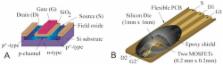
          The aims of this study were to characterize reinforced metal‐oxide‐semiconductor field‐effect transistor (MOSFET) dosimeters to assess the measurement uncertainty, single exposure low‐dose limit with acceptable accuracy, and the number of exposures required to attain the corresponding limit of the thermoluminescent dosimeters (TLD). The second aim was to characterize MOSFET dosimeter sensitivities for two dental photon energy ranges, dose dependency, dose rate dependency, and accumulated dose dependency. A further aim was to compare the performance of MOSFETs with those of TLDs in an anthropomorphic phantom head using a dentomaxillofacial CBCT device. The uncertainty was assessed by exposing 20 MOSFETs and a Barracuda MPD reference dosimeter. The MOSFET dosimeter sensitivities were evaluated for two photon energy ranges (50–90 kVp) using a constant dose and polymethylmethacrylate backscatter material. MOSFET and TLD comparative point‐dose measurements were performed on an anthropomorphic phantom that was exposed with a clinical CBCT protocol. The MOSFET single exposure low dose limit (25% uncertainty, k = 2 ) was 1.69 mGy. An averaging of eight MOSFET exposures was required to attain the corresponding TLD (0.3 mGy) low‐dose limit. The sensitivity was 3.09 ± 0.13   mV / mGy independently of the photon energy used. The MOSFET dosimeters did not present dose or dose rate sensitivity but, however, presented a 1% decrease of sensitivity per 1000 mV for accumulated threshold voltages between 8300 mV and 17500 mV. The point doses in an anthropomorphic phantom ranged for MOSFETs between 0.24 mGy and 2.29 mGy and for TLDs between 0.25 and 2.09 mGy, respectively. The mean difference was −8%. The MOSFET dosimeters presented statistically insignificant energy dependency. By averaging multiple exposures, the MOSFET dosimeters can achieve a TLD‐comparable low‐dose limit and constitute a feasible method for diagnostic dosimetry using anthropomorphic phantoms. However, for single in vivo measurements ( < 1.7   mGy ) the sensitivity is too low.

          PACS number: 87.50.wj

          Related collections

          Most cited references30

          • Record: found
          • Abstract: found
          • Article: not found

          Effective dose range for dental cone beam computed tomography scanners.

          To estimate the absorbed organ dose and effective dose for a wide range of cone beam computed tomography scanners, using different exposure protocols and geometries. Two Alderson Radiation Therapy anthropomorphic phantoms were loaded with LiF detectors (TLD-100 and TLD-100 H) which were evenly distributed throughout the head and neck, covering all radiosensitive organs. Measurements were performed on 14 CBCT devices: 3D Accuitomo 170, Galileos Comfort, i-CAT Next Generation, Iluma Elite, Kodak 9000 3D, Kodak 9500, NewTom VG, NewTom VGi, Pax-Uni3D, Picasso Trio, ProMax 3D, Scanora 3D, SkyView, Veraviewepocs 3D. Effective dose was calculated using the ICRP 103 (2007) tissue weighting factors. Effective dose ranged between 19 and 368 μSv. The largest contributions to the effective dose were from the remainder tissues (37%), salivary glands (24%), and thyroid gland (21%). For all organs, there was a wide range of measured values apparent, due to differences in exposure factors, diameter and height of the primary beam, and positioning of the beam relative to the radiosensitive organs. The effective dose for different CBCT devices showed a 20-fold range. The results show that a distinction is needed between small-, medium-, and large-field CBCT scanners and protocols, as they are applied to different indication groups, the dose received being strongly related to field size. Furthermore, the dose should always be considered relative to technical and diagnostic image quality, seeing that image quality requirements also differ for patient groups. The results from the current study indicate that the optimisation of dose should be performed by an appropriate selection of exposure parameters and field size, depending on the diagnostic requirements. Copyright © 2010 Elsevier Ireland Ltd. All rights reserved.
            Bookmark
            • Record: found
            • Abstract: found
            • Article: not found

            Dosimetry of two extraoral direct digital imaging devices: NewTom cone beam CT and Orthophos Plus DS panoramic unit.

            This study provides effective dose measurements for two extraoral direct digital imaging devices, the NewTom 9000 cone beam CT (CBCT) unit and the Orthophos Plus DS panoramic unit. Thermoluminescent dosemeters were placed at 20 sites throughout the layers of the head and neck of a tissue-equivalent RANDO phantom. Variations in phantom orientation and beam collimation were used to create three different CBCT examination techniques: a combined maxillary and mandibular scan (Max/Man), a maxillary scan and a mandibular scan. Ten exposures for each technique were used to ensure a reliable measure of radiation from the dosemeters. Average tissue-absorbed dose, weighted equivalent dose and effective dose were calculated for each major anatomical site. Effective doses of individual organs were summed with salivary gland exposures (E(SAL)) and without salivary gland exposures (E(ICRP60)) to calculate two measures of whole-body effective dose. The effective doses for CBCT were: Max/Man scan, E(ICRP60)=36.3 micro Sv, E(SAL)=77.9 micro Sv; maxillary scan, E(ICRP60)=19.9 micro Sv, E(SAL)=41.5 micro Sv; and mandibular scan, E(ICRP60)=34.7 micro Sv, E(SAL)=74.7 micro Sv. Effective doses for the panoramic examination were E(ICRP60)=6.2 micro Sv and E(SAL)=22.0 micro Sv. When viewed in the context of potential diagnostic yield, the E(ICRP60) of 36.3 micro Sv for the NewTom compares favourably with published effective doses for conventional CT (314 micro Sv) and film tomography (2-9 micro Sv per image). CBCT examinations resulted in doses that were 3-7 (E(ICRP60)) and 2-4 (E(SAL)) times the panoramic doses observed in this study.
              Bookmark
              • Record: found
              • Abstract: found
              • Article: not found

              Computation of bremsstrahlung X-ray spectra and comparison with spectra measured with a Ge(Li) detector.

              A method of computing theoretical X-ray spectra in the range 30-150 kV is presented. The theoretical spectra are compared with constant potential, high resolution spectra from a tungsten target measured with a Ge(Li) detector, for a range of target angles, tube voltage and filtrations. Above 100 kV the spectra were also measured with a NaI detector but, as there was good agreement between the Ge(Li) and NaI detectors, only the former are presented. Spectra computed using Kramers' theory are also included for comparison, giving fairly good agreement at large target angles (30 degrees) but becoming gradually worse as the target angle decreased. Spectra may be computed by this method for any desired filtration, target angle, and tube voltage between 30 and 150 kV, in excellent agreement with the measured data.
                Bookmark

                Author and article information

                Contributors
                juha.koivisto@planmeca.com
                Journal
                J Appl Clin Med Phys
                J Appl Clin Med Phys
                10.1002/(ISSN)1526-9914
                ACM2
                Journal of Applied Clinical Medical Physics
                John Wiley and Sons Inc. (Hoboken )
                1526-9914
                08 July 2015
                July 2015
                : 16
                : 4 ( doiID: 10.1002/acm2.2015.16.issue-4 )
                : 266-278
                Affiliations
                [ 1 ] Department of Physics University of Helsinki Helsinki Finland
                [ 2 ] Department of Oral and Maxillofacial Surgery/Pathology VU University Medical Center Amsterdam The Netherlands
                [ 3 ] International Docrates Cancer Center Helsinki Finland
                [ 4 ] Dental Diagnostic Center Freiburg Germany
                [ 5 ] HUS Helsinki Medical Imaging Center University of Helsinki Helsinki Finland
                Author notes
                [*] [* ] a Corresponding author: Juha H. Koivisto, Department of Physics, 1 University of Helsinki, Gustaf Hällströmin katu 2a, FI‐00560, Helsinki, Finland; phone: (358) 40 554 66 45; fax: (358) 20 7795 676; email: juha.koivisto@ 123456planmeca.com

                Article
                ACM20266
                10.1120/jacmp.v16i4.5433
                5690001
                26219008
                b8108215-2c7d-4103-8bf5-2c6b7404d0ac
                © 2015 The Authors.

                This is an open access article under the terms of the Creative Commons Attribution License, which permits use, distribution and reproduction in any medium, provided the original work is properly cited.

                History
                : 07 November 2014
                : 15 March 2015
                Page count
                Figures: 6, Tables: 2, References: 36, Pages: 13, Words: 5521
                Categories
                Radiation Measurements
                Radiation Measurements
                Custom metadata
                2.0
                acm20266
                July 2015
                Converter:WILEY_ML3GV2_TO_NLMPMC version:5.2.5 mode:remove_FC converted:16.11.2017

                mosfet dosimeter,x‐ray,radiation exposure
                mosfet dosimeter, x‐ray, radiation exposure

                Comments

                Comment on this article