10
views
0
recommends
+1 Recommend
0 collections
    0
    shares
      • Record: found
      • Abstract: found
      • Article: found
      Is Open Access

      Spinocerebellar ataxia: Functional analysis of the stomatognathic system

      research-article

      Read this article at

          There is no author summary for this article yet. Authors can add summaries to their articles on ScienceOpen to make them more accessible to a non-specialist audience.

          Abstract

          Background

          Neurodegenerative diseases that affect the cerebellum, especially in elderly individuals, cause impairment of motor coordination and quality of life. The presente study evaluated the electromyographic activity and thickness of the right and left masseter and temporal muscles, and the maximum molar bite force of individuals with spinocerebellar ataxia.

          Material and Methods

          Twenty-eight individuals were divided into two groups: those with (n=14) and without (n=14) spinocerebellar ataxia. Data on the masticatory muscles obtained from the electromyographic activity (resting, right and left laterality and protrusion), muscle thickness (maximal voluntary contraction and tensile strength) and maximum bite force (right and left) were tabulated and descriptive analysis using Student’s t-test ( P ≤ 0.05).

          Results

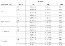
          In the comparison between groups, greater electromyographic activity was demonstrated for individuals with spinocerebellar ataxia, with a statistically significant difference in protrusion and laterality for the temporal muscles ( P = 0.05). There was no statistically significant difference between the groups for masticatory muscles thickness in the conditions evaluated. For maximum molar bite force, the group with spinocerebellar ataxia showed lower bite force ( P ≤ 0.05).

          Conclusions

          The data obtained suggest that spinocerebellar ataxia promotes functional reduction in the stomatognathic system, mainly affecting the electromyographic activity and bite force, hindering chewing, with a resultant alteration of nutritional intake and a decrease of quality of life.

          Key words:Spinocerebellar ataxia, electromyography, muscle thickness, bite force, masseter muscle, temporal muscle.

          Related collections

          Most cited references31

          • Record: found
          • Abstract: found
          • Article: not found

          Development of recommendations for SEMG sensors and sensor placement procedures.

          The knowledge of surface electromyography (SEMG) and the number of applications have increased considerably during the past ten years. However, most methodological developments have taken place locally, resulting in different methodologies among the different groups of users.A specific objective of the European concerted action SENIAM (surface EMG for a non-invasive assessment of muscles) was, besides creating more collaboration among the various European groups, to develop recommendations on sensors, sensor placement, signal processing and modeling. This paper will present the process and the results of the development of the recommendations for the SEMG sensors and sensor placement procedures. Execution of the SENIAM sensor tasks, in the period 1996-1999, has been handled in a number of partly parallel and partly sequential activities. A literature scan was carried out on the use of sensors and sensor placement procedures in European laboratories. In total, 144 peer-reviewed papers were scanned on the applied SEMG sensor properties and sensor placement procedures. This showed a large variability of methodology as well as a rather insufficient description. A special workshop provided an overview on the scientific and clinical knowledge of the effects of sensor properties and sensor placement procedures on the SEMG characteristics. Based on the inventory, the results of the topical workshop and generally accepted state-of-the-art knowledge, a first proposal for sensors and sensor placement procedures was defined. Besides containing a general procedure and recommendations for sensor placement, this was worked out in detail for 27 different muscles. This proposal was evaluated in several European laboratories with respect to technical and practical aspects and also sent to all members of the SENIAM club (>100 members) together with a questionnaire to obtain their comments. Based on this evaluation the final recommendations of SENIAM were made and published (SENIAM 8: European recommendations for surface electromyography, 1999), both as a booklet and as a CD-ROM. In this way a common body of knowledge has been created on SEMG sensors and sensor placement properties as well as practical guidelines for the proper use of SEMG.
            Bookmark
            • Record: found
            • Abstract: found
            • Article: not found

            Spinocerebellar ataxias: prospects and challenges for therapy development

            The spinocerebellar ataxias (SCAs) comprise more than 40 autosomal dominant neurodegenerative disorders that present principally with progressive ataxia. Within the past few years, studies of pathogenic mechanisms in the SCAs have led to the development of promising therapeutic strategies, especially for SCAs caused by polyglutamine-coding CAG repeats. Nucleotide-based gene-silencing approaches that target the first steps in the pathogenic cascade are one promising approach not only for polyglutamine SCAs but also for the many other SCAs caused by toxic mutant proteins or RNA. For these and other emerging therapeutic strategies, well-coordinated preparation is needed for fruitful clinical trials. To accomplish this goal, investigators from the United States and Europe are now collaborating to share data from their respective SCA cohorts. Increased knowledge of the natural history of SCAs, including of the premanifest and early symptomatic stages of disease, will improve the prospects for success in clinical trials of disease-modifying drugs. In addition, investigators are seeking validated clinical outcome measures that demonstrate responsiveness to changes in SCA populations. Findings suggest that MRI and magnetic resonance spectroscopy biomarkers will provide objective biological readouts of disease activity and progression, but more work is needed to establish disease-specific biomarkers that track target engagement in therapeutic trials. Together, these efforts suggest that the development of successful therapies for one or more SCAs is not far away.
              Bookmark
              • Record: found
              • Abstract: found
              • Article: not found

              Age and gender influence on maximal bite force and masticatory muscles thickness.

              The present study aimed investigate the age and gender influence on maximal molar bite force and at outlining the criteria for normal masticatory muscle development in a sample of 177 Brazilian Caucasian dentate individuals aged 7-80 years divided into five age groups: I (7-12 years), II (13-20 years), III (21-40 years), IV (41-60 years), and V (61-80 years). Except for Group V, which comprised nine women and eight men, all groups were equally divided in respect to gender (20 M/20 F). Bite force was recorded with a mouth-adapted 1000N dynamometer and the highest out of three records was regarded as the maximal bite force. The data were submitted to multivariate statistical analysis (SPSS 17.0 p<0.05). Effects of group and gender were found, but no interactions between them. The ANOVA showed significant differences between groups bilaterally. Bonferroni's test showed that group I had significantly lower bite force means at both sides as compared to all groups, except group V. No differences were found between the left and right sides. In all the groups, gender was found to be a significant factor associated with maximal bite force. A global comparison including all the subjects and measures showed that the means of men were approximately 30% higher than those of women. Within-group comparisons yielded similar results in all groups. Muscle thickness was measured with a SonoSite Titan ultrasound tool using a high-resolution real-time 56mm/10MHz linear-array transducer. Three ultrasound images were obtained from the bilateral masseter and temporal muscles at rest and at maximal voluntary contraction. The means of the three measures in each clinical condition were analyzed with multivariate statistical analysis (SPSS 17.0 p<0.05). A gradual increase in thickness of the masseter and temporal muscles was found both at rest and maximal voluntary contraction for groups I to IV, whereas a decrease in muscle thickness was observed in group V. Multivariate analysis showed that in both conditions there was an effect of group and gender. The study of the development of the stomatognathic system in relation to age and gender can provide useful data for the identification of normal and impaired functioning patterns. The results of this study indicate that age and gender are associated with structural and functional alterations in the muscles of the stomatognathic system. Copyright 2010 Elsevier Ltd. All rights reserved.
                Bookmark

                Author and article information

                Journal
                Med Oral Patol Oral Cir Bucal
                Med Oral Patol Oral Cir Bucal
                Medicina Oral S.L.
                Medicina Oral, Patología Oral y Cirugía Bucal
                Medicina Oral S.L.
                1698-4447
                1698-6946
                March 2019
                : 24
                : 2
                : e165-e171
                Affiliations
                [1 ]DDS, Professor. School of Dentistry of Ribeirão Preto, University of São Paulo Department of Morphology, Physiology and Basic Pathology. Rehabilitation and Ribeirão Preto Medical School, University of São Paulo, São Paulo, Brazil
                [2 ]DDS, PhD, Professor. School of Dentistry of Ribeirão Preto, University of São Paulo Department of Morphology, Physiology and Basic Pathology. National Institute and Technology - Translational Medicine (INCT.TM). Postgraduate of the Faculty Anhanguera, Ribeirão Preto, São Paulo, Brazil
                [3 ]DDS, PhD. School of Dentistry of Ribeirão Preto, University of São Paulo Department of Morphology, Physiology and Basic Pathology, São Paulo, Brazil
                [4 ]DDS, PhD, Professor. School of Dentistry of Ribeirão Preto, University of São Paulo Department of Morphology, Physiology and Basic Pathology. Rehabilitation and Ribeirão Preto Medical School, University of São Paulo, São Paulo, Brazil
                [5 ]DDS. School of Dentistry of Ribeirão Preto, University of São Paulo Department of Morphology, Physiology and Basic Pathology, São Paulo, Brazil
                [6 ]DDS, PhD, Professor. Rehabilitation and Ribeirão Preto Medical School, University of São Paulo, São Paulo, Brazil
                [7 ]DDS, PhD, Professor. National Institute and Technology - Translational Medicine (INCT.TM). Rehabilitation and Ribeirão Preto Medical School, University of São Paulo, São Paulo, Brazil
                [8 ]DDS, PhD, Professor. School of Dentistry of Ribeirão Preto, University of São Paulo Department of Morphology, Physiology and Basic Pathology. National Institute and Technology - Translational Medicine (INCT.TM), São Paulo, Brazil
                Author notes
                School of Dentistry of Ribeirão Preto University of São Paulo. Avenida do Café, s/n Bairro Monte Alegre, CEP 14040-904 Ribeirão Preto SP, Brazil , E-mail: palinkas@ 123456usp.br

                Conflict of interest statement:All authors declare no conflict of interest.

                Article
                22839
                10.4317/medoral.22839
                6441597
                30818308
                3731b2fd-8724-4a18-b63c-400e425a8a67
                Copyright: © 2019 Medicina Oral S.L.

                This is an open-access article distributed under the terms of the Creative Commons Attribution License, which permits unrestricted use, distribution, and reproduction in any medium, provided the original work is properly cited.

                History
                : 4 February 2019
                : 27 October 2018
                Categories
                Research
                Oral Medicine and Pathology

                Surgery
                Surgery

                Comments

                Comment on this article