5
views
0
recommends
+1 Recommend
0 collections
    0
    shares
      • Record: found
      • Abstract: found
      • Article: found
      Is Open Access

      Across the spectrum: integrating multidimensional metal analytics for in situ metallomic imaging

      review-article
      a , b
      Metallomics
      Royal Society of Chemistry

      Read this article at

          There is no author summary for this article yet. Authors can add summaries to their articles on ScienceOpen to make them more accessible to a non-specialist audience.

          Abstract

          Taking a systems analytical approach to systems biology questions requires a network of multidimensional analytical tools to illuminate the many different functional and structural aspects of metals in biology.

          Abstract

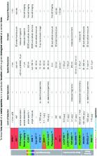
          To know how much of a metal species is in a particular location within a biological context at any given time is essential for understanding the intricate roles of metals in biology and is the fundamental question upon which the field of metallomics was born. Simply put, seeing is powerful. With the combination of spectroscopy and microscopy, we can now see metals within complex biological matrices complemented by information about associated molecules and their structures. With the addition of mass spectrometry and particle beam based techniques, the field of view grows to cover greater sensitivities and spatial resolutions, addressing structural, functional and quantitative metallomic questions from the atomic level to whole body processes. In this perspective, I present a paradigm shift in the way we relate to and integrate current and developing metallomic analytics, highlighting both familiar and perhaps less well-known state of the art techniques for in situ metallomic imaging, specific biological applications, and their use in correlative studies. There is a genuine need to abandon scientific silos and, through the establishment of a metallomic scientific platform for further development of multidimensional analytics for in situ metallomic imaging, we have an incredible opportunity to enhance the field of metallomics and demonstrate how discovery research can be done more effectively.

          Related collections

          Most cited references16

          • Record: found
          • Abstract: found
          • Article: found
          Is Open Access

          An introduction to optical super-resolution microscopy for the adventurous biologist

            Bookmark
            • Record: found
            • Abstract: found
            • Article: not found

            Simultaneous cryo X-ray ptychographic and fluorescence microscopy of green algae.

            Trace metals play important roles in normal and in disease-causing biological functions. X-ray fluorescence microscopy reveals trace elements with no dependence on binding affinities (unlike with visible light fluorophores) and with improved sensitivity relative to electron probes. However, X-ray fluorescence is not very sensitive for showing the light elements that comprise the majority of cellular material. Here we show that X-ray ptychography can be combined with fluorescence to image both cellular structure and trace element distribution in frozen-hydrated cells at cryogenic temperatures, with high structural and chemical fidelity. Ptychographic reconstruction algorithms deliver phase and absorption contrast images at a resolution beyond that of the illuminating lens or beam size. Using 5.2-keV X-rays, we have obtained sub-30-nm resolution structural images and ∼90-nm-resolution fluorescence images of several elements in frozen-hydrated green algae. This combined approach offers a way to study the role of trace elements in their structural context.
              Bookmark
              • Record: found
              • Abstract: found
              • Article: not found

              Golgi in copper homeostasis: a view from the membrane trafficking field.

              Copper is essential for a variety of important biological processes as a cofactor and regulator of many enzymes. Incorporation of copper into the secreted and plasma membrane-targeted cuproenzymes takes place in Golgi, a compartment central for normal copper homeostasis. The Golgi complex harbors copper-transporting ATPases, ATP7A and ATP7B that transfer copper from the cytosol into Golgi lumen for incorporation into copper-dependent enzymes. The Golgi complex also sends these ATPases to appropriate post-Golgi destinations to ensure correct Cu fluxes in the body and to avoid potentially toxic copper accumulation. Mutations in ATP7A or ATP7B or in the proteins that regulate their trafficking affect their exit from Golgi or subsequent retrieval to this organelle. This, in turn, disrupts the homeostatic Cu balance, resulting in copper deficiency (Menkes disease) or copper overload (Wilson disease). Research over the last decade has yielded significant insights into the enzymatic properties and cell biology of the copper ATPases. However, the mechanisms through which the Golgi regulates trafficking of ATP7A/7B and, therefore, maintains Cu homeostasis remain unclear. This review summarizes current data on the role of the Golgi in Cu metabolism and outlines questions and challenges that should be addressed to understand ATP7A and ATP7B trafficking mechanisms in health and disease.
                Bookmark

                Author and article information

                Journal
                Metallomics
                Metallomics
                Metallomics
                Royal Society of Chemistry
                1756-5901
                1756-591X
                1 January 2019
                30 November 2018
                : 11
                : 1
                : 29-49
                Affiliations
                [a ] King's College London , Mass Spectrometry , London Metallomics Facility , 4th Floor Franklin-Wilkins Building , 150 Stamford St. , London SE1 9NH , UK . Email: theodora.stewart@ 123456kcl.ac.uk
                [b ] King's College London , School of Biomedical Engineering and Imaging Sciences , 4th Floor Lambeth Wing, St. Thomas’ Hospital , London SE1 7EH , UK
                Author information
                http://orcid.org/0000-0002-5651-8664
                Article
                c8mt00235e
                10.1039/c8mt00235e
                6350628
                30499574
                2e7e0adf-5510-4545-879f-c797a18dc7ab
                This journal is © The Royal Society of Chemistry 2019

                This article is freely available. This article is licensed under a Creative Commons Attribution 3.0 Unported Licence (CC BY 3.0)

                History
                : 10 August 2018
                : 17 October 2018
                Categories
                Chemistry

                Comments

                Comment on this article