7
views
0
recommends
+1 Recommend
0 collections
    0
    shares
      • Record: found
      • Abstract: found
      • Article: found
      Is Open Access

      Combined Perfusion and Permeability Imaging Reveals Different Pathophysiologic Tissue Responses After Successful Thrombectomy

      research-article

      Read this article at

          There is no author summary for this article yet. Authors can add summaries to their articles on ScienceOpen to make them more accessible to a non-specialist audience.

          Abstract

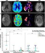
          Despite successful recanalization of large-vessel occlusions in acute ischemic stroke, individual patients profit to a varying degree. Dynamic susceptibility-weighted perfusion and dynamic T1-weighted contrast-enhanced blood-brain barrier permeability imaging may help to determine secondary stroke injury and predict clinical outcome. We prospectively performed perfusion and permeability imaging in 38 patients within 24 h after successful mechanical thrombectomy of an occlusion of the middle cerebral artery M1 segment. Perfusion alterations were evaluated on cerebral blood flow maps, blood-brain barrier disruption (BBBD) visually and quantitatively on k trans maps and hemorrhagic transformation on susceptibility-weighted images. Visual BBBD within the DWI lesion corresponded to a median k trans elevation (IQR) of 0.77 (0.41–1.4) min −1 and was found in all 7 cases of hypoperfusion (100%), in 10 of 16 cases of hyperperfusion (63%), and in only three of 13 cases with unaffected perfusion (23%). BBBD was significantly associated with hemorrhagic transformation ( p < 0.001). While BBBD alone was not a predictor of clinical outcome at 3 months (positive predictive value (PPV) = 0.8 [0.56–0.94]), hypoperfusion occurred more often in patients with unfavorable clinical outcome (PPV = 0.43 [0.10–0.82]) compared to hyperperfusion (PPV = 0.93 [0.68–1.0]) or unaffected perfusion (PPV = 1.0 [0.75–1.0]). We show that combined perfusion and permeability imaging reveals distinct infarct signatures after recanalization, indicating the severity of prior ischemic damage. It assists in predicting clinical outcome and may identify patients at risk of stroke progression.

          Supplementary Information

          The online version contains supplementary material available at 10.1007/s12975-020-00885-y.

          Related collections

          Most cited references45

          • Record: found
          • Abstract: found
          • Article: not found

          User-guided 3D active contour segmentation of anatomical structures: significantly improved efficiency and reliability.

          Active contour segmentation and its robust implementation using level set methods are well-established theoretical approaches that have been studied thoroughly in the image analysis literature. Despite the existence of these powerful segmentation methods, the needs of clinical research continue to be fulfilled, to a large extent, using slice-by-slice manual tracing. To bridge the gap between methodological advances and clinical routine, we developed an open source application called ITK-SNAP, which is intended to make level set segmentation easily accessible to a wide range of users, including those with little or no mathematical expertise. This paper describes the methods and software engineering philosophy behind this new tool and provides the results of validation experiments performed in the context of an ongoing child autism neuroimaging study. The validation establishes SNAP intrarater and interrater reliability and overlap error statistics for the caudate nucleus and finds that SNAP is a highly reliable and efficient alternative to manual tracing. Analogous results for lateral ventricle segmentation are provided.
            Bookmark
            • Record: found
            • Abstract: not found
            • Article: not found

            Improved Optimization for the Robust and Accurate Linear Registration and Motion Correction of Brain Images

              Bookmark
              • Record: found
              • Abstract: found
              • Article: not found

              A randomized trial of intraarterial treatment for acute ischemic stroke.

              In patients with acute ischemic stroke caused by a proximal intracranial arterial occlusion, intraarterial treatment is highly effective for emergency revascularization. However, proof of a beneficial effect on functional outcome is lacking. We randomly assigned eligible patients to either intraarterial treatment plus usual care or usual care alone. Eligible patients had a proximal arterial occlusion in the anterior cerebral circulation that was confirmed on vessel imaging and that could be treated intraarterially within 6 hours after symptom onset. The primary outcome was the modified Rankin scale score at 90 days; this categorical scale measures functional outcome, with scores ranging from 0 (no symptoms) to 6 (death). The treatment effect was estimated with ordinal logistic regression as a common odds ratio, adjusted for prespecified prognostic factors. The adjusted common odds ratio measured the likelihood that intraarterial treatment would lead to lower modified Rankin scores, as compared with usual care alone (shift analysis). We enrolled 500 patients at 16 medical centers in The Netherlands (233 assigned to intraarterial treatment and 267 to usual care alone). The mean age was 65 years (range, 23 to 96), and 445 patients (89.0%) were treated with intravenous alteplase before randomization. Retrievable stents were used in 190 of the 233 patients (81.5%) assigned to intraarterial treatment. The adjusted common odds ratio was 1.67 (95% confidence interval [CI], 1.21 to 2.30). There was an absolute difference of 13.5 percentage points (95% CI, 5.9 to 21.2) in the rate of functional independence (modified Rankin score, 0 to 2) in favor of the intervention (32.6% vs. 19.1%). There were no significant differences in mortality or the occurrence of symptomatic intracerebral hemorrhage. In patients with acute ischemic stroke caused by a proximal intracranial occlusion of the anterior circulation, intraarterial treatment administered within 6 hours after stroke onset was effective and safe. (Funded by the Dutch Heart Foundation and others; MR CLEAN Netherlands Trial Registry number, NTR1804, and Current Controlled Trials number, ISRCTN10888758.).
                Bookmark

                Author and article information

                Contributors
                angelika.hoffmann@med.uni-heidelberg.de
                Journal
                Transl Stroke Res
                Transl Stroke Res
                Translational Stroke Research
                Springer US (New York )
                1868-4483
                1868-601X
                11 January 2021
                11 January 2021
                2021
                : 12
                : 5
                : 799-807
                Affiliations
                [1 ]GRID grid.5253.1, ISNI 0000 0001 0328 4908, Department of Neuroradiology, , Heidelberg University Hospital, ; INF 400, 69120 Heidelberg, Germany
                [2 ]GRID grid.5253.1, ISNI 0000 0001 0328 4908, Department of Neurology, , Heidelberg University Hospital, ; Heidelberg, Germany
                [3 ]GRID grid.5253.1, ISNI 0000 0001 0328 4908, Division of Experimental Radiology, Department of Neuroradiology, , Heidelberg University Hospital, ; Heidelberg, Germany
                [4 ]GRID grid.8379.5, ISNI 0000 0001 1958 8658, Department of Neuroradiology, , Würzburg University Hospital, ; Würzburg, Germany
                [5 ]GRID grid.5734.5, ISNI 0000 0001 0726 5157, University Institute of Diagnostic and Interventional Neuroradiology, University Hospital Bern, Inselspital, , University of Bern, ; Bern, Switzerland
                Author information
                http://orcid.org/0000-0003-4552-0694
                Article
                885
                10.1007/s12975-020-00885-y
                8421283
                33432454
                edbe4650-eb59-4049-b009-e09dc3f44deb
                © The Author(s) 2021

                Open Access This article is licensed under a Creative Commons Attribution 4.0 International License, which permits use, sharing, adaptation, distribution and reproduction in any medium or format, as long as you give appropriate credit to the original author(s) and the source, provide a link to the Creative Commons licence, and indicate if changes were made. The images or other third party material in this article are included in the article's Creative Commons licence, unless indicated otherwise in a credit line to the material. If material is not included in the article's Creative Commons licence and your intended use is not permitted by statutory regulation or exceeds the permitted use, you will need to obtain permission directly from the copyright holder. To view a copy of this licence, visit http://creativecommons.org/licenses/by/4.0/.

                History
                : 22 August 2020
                : 10 December 2020
                : 28 December 2020
                Funding
                Funded by: FundRef http://dx.doi.org/10.13039/100009139, Deutsches Zentrum für Infektionsforschung;
                Award ID: Clinical Leave Stipend
                Award Recipient :
                Funded by: Guerbet
                Award ID: This research was supported by a funding of Guerbet, BP 57400, 95943 ROISSY CDG, FRANCE. The funders had no role in study design, data collection and analysis, decision to publish, or preparation of the manuscript.
                Categories
                Original Article
                Custom metadata
                © Springer Science+Business Media, LLC, part of Springer Nature 2021

                Cardiovascular Medicine
                permeability imaging,perfusion imaging,mechanical thrombectomy,secondary stroke injury,hyperperfusion

                Comments

                Comment on this article