1
views
0
recommends
+1 Recommend
0 collections
    0
    shares
      • Record: found
      • Abstract: found
      • Article: found
      Is Open Access

      Exceedingly rare incidence of a double inferior vena cava (IVC) with azygos continuation of left IVC

      case-report

      Read this article at

          There is no author summary for this article yet. Authors can add summaries to their articles on ScienceOpen to make them more accessible to a non-specialist audience.

          Abstract

          Key Clinical Message

          Because of the complex embryonic origin of the abdominal venous structures, IVC and azygous systems can show numerous and even previously unreported anatomical variations and anomalies. Also, evaluating major vascular structures should not be dismissed in non‐contrast‐enhanced CT as it can provide valuable information about these structures.

          Abstract

          Double IVC is a rare occurrence of IVC anatomical variations and congenital anomalies. Herein, we discuss a case of a very rare type of double IVC that has not been reported in the literature before. A non‐contrast‐enhanced CT study was performed for a 34‐year‐old patient who visited our ER to evaluate for urolithiasis, during which two IVCs were noted. Each renal vein joined the ipsilateral IVC at a perpendicular angle. Unusually, the right IVC was formed from the confluence of both left and right common iliac veins (CIV), and the left IVC—Instead of crossing the midline at the renal veins level and reuniting the right IVC—cranially contributed to the azygos vein formation and caudally joined the left CIV. Also, there were some small communicating veins between the two IVCs and the left gonadal vein was slightly dilated before suggesting a reflux from the left renal vein (LRV). A complimentary doppler ultrasound exam confirmed the diagnosis and revealed a left‐side varicocele. Although rare cases of hemiazygos continuation and interiliac connections of left‐side IVC in the cases of double‐IVC have been reported previously, a complete confluence of CIVs is rare. The main differential diagnosis is retro‐aortic left renal vein (RLRV) type IV which seems to have an oblique course. Radiologists and surgeons should expect previously unreported variations in the vena cava system. Furthermore, reviewing the main abdominal vasculature should not be dismissed in non‐contrast CT exams.

          Abstract

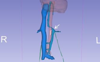
          The operator assisted 3D reconstruction of the main retroperitoneal vasculature: the Aorta is shown in red; the right renal vein, right Inferior Vena Cava (IVC), and common iliac veins are depicted in dark blue; the left renal vein, left IVC, azygos vein, and accessory hemiazygos vein are illustrated in cyan; and the gonadal veins are represented in green. The left IVC exhibits multiple connections to the right IVC and the left Common Iliac Vein (CIV) caudally. It follows a vertical path and intersects with the left renal vein at a right angle. The superior segment of the left IVC, (arrow) does not merge with the right IVC after crossing the midline above the confluence of the left renal vein. Instead, it contributes to the formation of the azygos vein at the T12 vertebral level.

          Related collections

          Most cited references18

          • Record: found
          • Abstract: found
          • Article: not found

          Spectrum of congenital anomalies of the inferior vena cava: cross-sectional imaging findings.

          Congenital anomalies of the inferior vena cava (IVC) and its tributaries have become more commonly recognized in asymptomatic patients. The embryogenesis of the IVC is a complex process involving the formation of several anastomoses between three paired embryonic veins. The result is numerous variations in the basic venous plan of the abdomen and pelvis. A left IVC typically ends at the left renal vein, which crosses anterior to the aorta to form a normal right-sided prerenal IVC. In double IVC, the left IVC typically ends at the left renal vein, which crosses anterior to the aorta to join the right IVC. In azygos continuation of the IVC, the prerenal IVC passes posterior to the diaphragmatic crura to enter the thorax as the azygos vein. In circumaortic left renal vein, one left renal vein crosses anterior to the aorta and another crosses posterior to the aorta. In retroaortic left renal vein, the left renal vein passes posterior to the aorta. In circumcaval ureter, the proximal ureter courses posterior to the IVC. Other anomalies include absence of the infrarenal IVC or the entire IVC. These anomalies can have significant clinical implications. Awareness of these anomalies is necessary to avoid diagnostic pitfalls.
            Bookmark
            • Record: found
            • Abstract: found
            • Article: not found

            Inferior vena cava anomalies and variations: imaging and rare clinical findings

            Purpose The aim of this paper is to summarize imaging findings of some frequent and infrequent inferior vena cava (IVC) anomalies and variations. Conclusions IVC anomalies should be suspected in patients presenting with pulmonary emboli, chronic pain, and deep vein thrombosis. To correctly characterize and classify IVC anomalies and variations is of crucial importance for proper planning of surgical interventions and thus for avoiding serious complications. Key Points • IVC anomalies should be suspected in patients with pulmonary emboli, pain, and venous thrombosis. • Awareness of IVC anomalies and variations is crucial for clinical and surgical procedures. • Unawareness of these anomalies may lead to severe and deadly complications.
              Bookmark
              • Record: found
              • Abstract: found
              • Article: not found

              Retroaortic left renal vein: multidetector computed tomography angiography findings and its clinical importance.

              To evaluate the appearance, type, frequency, and clinical importance of retroaortic left renal vein (RLRV) in patients examined with multidetector computed tomography (MDCT) angiography. A total of 1856 patients who underwent CT with urological symptoms (hematuria, flank and abdominal pain, left gonadal vein varicocele) (n = 889) or with symptoms other than urological ones (n = 967) were prospectively evaluated for the presence of RLRV. CT was performed with 110 ml of iodinated contrast material through the antecubital vein at a rate of 3.5 ml/s. Late arterial and early venous phase volumetric data sets were acquired at 30 and 65 s, respectively, from the start of the intravenous injection of contrast medium. In addition to axial images, multiplanar reconstructions (MPR), maximum-intensity projection (MIP), and three-dimensional volume-rendering (3D VR) images were used to assess left renal vein anomalies. Left renal vein anomalies were classified into four types according to their appearance: I) RLRV joining the inferior vena cava (IVC) in the orthotopic position; II) RLRV joining the IVC at level L4-L5; III) circumaortic or collar left renal vein; IV) RLRV joining the left common iliac vein. RLRV was detected in 68 (3.6%) of the 1856 patients, with 26, 22, 17, and three of types I, II, III, and IV, respectively. Forty-four of the 68 patients with RLRV (65%) were in the group with urological symptoms, while 24 patients (35%) were in the group without urological symptoms. Compression of the RLRV was found in 16 patients in the urological symptoms group, while compression was detected in only three patients in the other group. This difference was statistically significant (P<0.05). The most common urological symptom was hematuria. The frequency of urological symptoms was higher in groups II and IV compared to the other groups. MDCT angiography with axial, MPR, MIP, and 3D VR images is effective in the detection of vascular renal anomalies such as RLRV. Diagnosing RLRV and differentiating it from other pathologic conditions causing hematuria is important in order to avoid complications during retroperitoneal surgery or interventional procedures.
                Bookmark

                Author and article information

                Contributors
                saeedakbarzadehpasha@gmail.com
                Journal
                Clin Case Rep
                Clin Case Rep
                10.1002/(ISSN)2050-0904
                CCR3
                Clinical Case Reports
                John Wiley and Sons Inc. (Hoboken )
                2050-0904
                23 May 2024
                June 2024
                : 12
                : 6 ( doiID: 10.1002/ccr3.v12.6 )
                : e8981
                Affiliations
                [ 1 ] Department of Radiology, School of Medicine Iran University of Medical Sciences Tehran Iran
                Author notes
                [*] [* ] Correspondence

                Saeed Akbarzadeh pasha, Department of Radiology, School of Medicine, Iran University of Medical Sciences, Hasheminejd Kidney Center, Vali‐nejad Str., Vanak Sq. Vali‐e‐asr Boul, Tehran, Iran.

                Email: saeedakbarzadehpasha@ 123456gmail.com

                Author information
                https://orcid.org/0000-0003-4669-5330
                https://orcid.org/0000-0003-2839-9861
                https://orcid.org/0000-0003-4599-999X
                Article
                CCR38981 CCR3-2024-01-0036.R1
                10.1002/ccr3.8981
                11116462
                a31e2dc4-28cf-48cc-943c-f4b1af88ba36
                © 2024 The Author(s). Clinical Case Reports published by John Wiley & Sons Ltd.

                This is an open access article under the terms of the http://creativecommons.org/licenses/by/4.0/ License, which permits use, distribution and reproduction in any medium, provided the original work is properly cited.

                History
                : 23 April 2024
                : 30 January 2024
                : 13 May 2024
                Page count
                Figures: 6, Tables: 1, Pages: 7, Words: 3900
                Categories
                Urology/Andrology
                Case Report
                Case Report
                Custom metadata
                2.0
                June 2024
                Converter:WILEY_ML3GV2_TO_JATSPMC version:6.4.3 mode:remove_FC converted:24.05.2024

                azygos continuation of left ivc,case report,double inferior vena cava,left renal vein,left‐side inferior vena cava

                Comments

                Comment on this article