26
views
0
recommends
+1 Recommend
0 collections
    0
    shares
      • Record: found
      • Abstract: found
      • Article: found
      Is Open Access

      Tumor budding, a novel prognostic indicator for predicting stage progression in T1 bladder cancers

      research-article

      Read this article at

          There is no author summary for this article yet. Authors can add summaries to their articles on ScienceOpen to make them more accessible to a non-specialist audience.

          Abstract

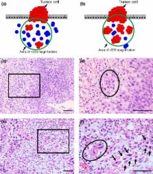
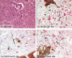
          Tumor budding has been defined as an isolated single cancer cell or a cluster composed of fewer than five cancer cells scattered in the stroma. It is a strong predictor for lymph node metastasis in T1 colorectal cancer. We introduced this concept to T1 non‐muscle invasive bladder cancer and evaluated whether tumor budding could have a prognostic impact on the clinical outcome. We identified 121 consecutive patients with newly diagnosed T1 bladder cancer between 1994 and 2014 at Keio University Hospital. All slides were re‐reviewed by a dedicated uropathologist. Budding foci were counted under ×200 magnification. When the number of budding foci was 10 or more, tumor budding was defined as positive. The relationship between tumor budding and clinical outcomes was assessed using a multivariate analysis. The median follow‐up was 52 months. Tumor budding was positive in 21 patients (17.4%). Tumor budding was significantly associated with T1 substaging, tumor architecture and lymphovascular invasion. The 5‐year progression‐free survival rate in T1 bladder cancer patients with tumor budding was 53.8%, which was significantly lower than that in patients without tumor budding (88.4%, P = 0.001). A multivariate Cox regression analysis revealed that tumor budding was independently associated with stage progression ( P = 0.002, hazard ratio = 4.90). In a subgroup of patients treated with bacillus Calmette‐Guérin instillation ( n = 88), tumor budding was also independently associated with stage progression ( P = 0.003, hazard ratio = 5.65). Tumor budding may be a novel indicator for predicting stage progression in T1 bladder cancer, and would likely be easily introduced in clinical practice.

          Related collections

          Most cited references23

          • Record: found
          • Abstract: found
          • Article: not found

          Japanese Society for Cancer of the Colon and Rectum (JSCCR) Guidelines 2014 for treatment of colorectal cancer

          Colorectal cancer is a major cause of death in Japan, where it accounts for the largest number of deaths from malignant neoplasms among women and the third largest number among men. Many new methods of treatment have been developed during recent decades. The Japanese Society for Cancer of the Colon and Rectum Guidelines 2014 for treatment of colorectal cancer (JSCCR Guidelines 2014) have been prepared as standard treatment strategies for colorectal cancer, to eliminate treatment disparities among institutions, to eliminate unnecessary treatment and insufficient treatment, and to deepen mutual understanding among health-care professionals and patients by making these guidelines available to the general public. These guidelines have been prepared as a result of consensuses reached by the JSCCR Guideline Committee on the basis of careful review of evidence retrieved by literature searches and taking into consideration the medical health insurance system and actual clinical practice in Japan. They can, therefore, be used as a guide for treating colorectal cancer in clinical practice. More specifically, they can be used as a guide to obtaining informed consent from patients and choosing the method of treatment for each patient. As a result of the discussions of the Guideline Committee, controversial issues were selected as clinical questions, and recommendations were made. Each recommendation is accompanied by a classification of the evidence and a classification of recommendation categories, on the basis of consensus reached by Guideline Committee members. Here we present the English version of the JSCCR Guidelines 2014.
            Bookmark
            • Record: found
            • Abstract: found
            • Article: not found

            Risk factors for an adverse outcome in early invasive colorectal carcinoma.

            Various histologic findings exist for managing patients with malignant polyps. Our goal was to determine the criteria for a conservative approach to patients with locally excised early invasive carcinoma. In 292 early invasive tumors (local resection followed by laparotomy [80 tumors, group A], local resection only [41 tumors, group B], and primarily laparotomy [171 tumors, group C], potential parameters for nodal involvement were analyzed. The status of the endoscopic resection margin also was examined for the risk for intramural residual tumor. Unfavorable tumor grade, definite vascular invasion, and tumor budding were the combination of qualitative factors that most effectively discriminated the risk for nodal involvement in patients in groups A-C. The nodal involvement rate was 0.7%, 20.7%, and 36.4% in the no-risk, single-risk, and multiple-risks group, respectively. Thirty-two and 9 patients from group B were assigned to the no-risk and one-risk group, respectively; extramural recurrence occurred in 2 patients with risk factors. Considering quantitative risk parameters for submucosal invasion (i.e., width > or =4000 microm or depth > or =2000 microm), nodal involvement (including micrometastases) was not observed in the redefined no-risk group that accounted for about 25% of the patients from groups A and C. An insufficiency of endoscopic resection could be evaluated most precisely based on the coagulation-involving tumor, rather than the 1-mm rule for the resection margin. Provided that the criterion of sufficient excision is satisfied, the absence of an unfavorable tumor grade, vascular invasion, tumor budding, and extensive submucosal invasion would be the strict criteria for a wait-and-see policy.
              Bookmark
              • Record: found
              • Abstract: found
              • Article: found
              Is Open Access

              Epithelial mesenchymal transition and tumor budding in aggressive colorectal cancer: Tumor budding as oncotarget

              Epithelial mesenchymal transition (EMT) is proposed as a critical mechanism for the acquisition of malignant phenotypes by epithelial cells. In colorectal cancer, tumor cells having undergone EMT are histologically represented by the presence of tumor buds defined as single cells or small clusters of de-differentiated tumor cells at the invasive front. Tumor budding is not a static, histological feature rather it represents a snap-shot of a dynamic process undertaken by an aggressive tumor with the potential to disseminate and metastasize. Strong, consistent evidence shows that tumor budding is a predictor of lymph node metastasis, distant metastatic disease, local recurrence, worse overall and disease-free survival time and an independent prognostic factor. Moreover, the International Union against Cancer (UICC) recognizes tumor budding as a highly relevant, additional prognostic parameter. The aim of this review is to summarize the evidence supporting the implementation of tumor budding into diagnostic pathology and patient management and additionally to illustrate its worthiness as a potential therapeutic target.
                Bookmark

                Author and article information

                Contributors
                eiji-k@kb3.so-net.ne.jp
                Journal
                Cancer Sci
                Cancer Sci
                10.1111/(ISSN)1349-7006
                CAS
                Cancer Science
                John Wiley and Sons Inc. (Hoboken )
                1347-9032
                1349-7006
                12 August 2016
                September 2016
                : 107
                : 9 ( doiID: 10.1111/cas.2016.107.issue-9 )
                : 1338-1344
                Affiliations
                [ 1 ] Department of UrologyKeio University School of Medicine TokyoJapan
                [ 2 ] Division of Diagnostic PathologyKeio University School of Medicine TokyoJapan
                Author notes
                [*] [* ] Correspondence

                Eiji Kikuchi, Department of Urology, Keio University School of Medicine, 35 Shinanomachi, Shinjuku‐ku, Tokyo 160‐0016, Japan.

                Tel: +81‐3‐5363‐3825; Fax: +81‐3‐3225‐1985;

                E‐mail: eiji-k@ 123456kb3.so-net.ne.jp

                Article
                CAS12990
                10.1111/cas.12990
                5021027
                27317460
                549fd13c-2946-4481-b0bc-e23183937b6f
                © 2016 The Authors. Cancer Science published by John Wiley & Sons Australia, Ltd on behalf of Japanese Cancer Association.

                This is an open access article under the terms of the Creative Commons Attribution‐NonCommercial‐NoDerivs License, which permits use and distribution in any medium, provided the original work is properly cited, the use is non‐commercial and no modifications or adaptations are made.

                History
                : 01 March 2016
                : 07 June 2016
                : 16 June 2016
                Page count
                Figures: 2, Tables: 3, Pages: 7, Words: 5884
                Funding
                Funded by: Keio University Grant‐in‐Aid for Encouragement of Young Medical Scientists
                Award ID: 02‐002‐0027
                Funded by: Japan Society for the Promotion of Science KAKENHI
                Award ID: 15K20107
                Categories
                Original Article
                Original Articles
                Pathology
                Custom metadata
                2.0
                cas12990
                September 2016
                Converter:WILEY_ML3GV2_TO_NLMPMC version:4.9.4 mode:remove_FC converted:13.09.2016

                Oncology & Radiotherapy
                disease progression,pathology,recurrence,transitional cell,urinary bladder neoplasms

                Comments

                Comment on this article