2
views
0
recommends
+1 Recommend
0 collections
    0
    shares
      • Record: found
      • Abstract: found
      • Article: found
      Is Open Access

      Analysis of blood group antigens on MUC5AC in mucinous ovarian cancer tissues using in situ proximity ligation assay

      research-article

      Read this article at

          There is no author summary for this article yet. Authors can add summaries to their articles on ScienceOpen to make them more accessible to a non-specialist audience.

          Abstract

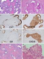
          MUC5AC has been indicated to be a marker for mucinous ovarian cancer (OC). We investigated the use of in situ proximity ligation assay (PLA) for blood group ABH expressing MUC5AC to differentiate between serous and mucinous OC, to validate preceding observations that also MUC5AC ABH expression is increased in mucinous OC. We developed PLA for anti-A, B, and H/anti-MUC5AC and a PLA using a combined lectin Ulex europaeus agglutinin I (UEA I)/anti-MUC5AC assay. The PLAs were verified with mass spectrometry, where mucinous OC secretor positive patients’ cysts fluids containing ABH O-linked oligosaccharides also showed positive OC tissue PLA staining. A nonsecretor mucinous OC cyst fluid was negative for ABH and displayed negative PLA staining of the matched tissue. Using the UEA I/MUC5AC PLA, we screened a tissue micro array of 410 ovarian tissue samples from patients with various stages of mucinous or serous OC, 32 samples with metastasis to the ovaries and 34 controls. The PLA allowed differentiating mucinous tumors with a sensitivity of 84% and a specificity of 97% both against serous cancer but also compared to tissues from controls. This sensitivity is close to the expected incidence of secretor individuals in a population. The recorded sensitivity was also found to be higher compared to mucinous type cancer with metastasis to the ovaries, where only 32% were positive. We conclude that UEA 1/MUC5AC PLA allows glycospecific differentiation between serous and mucinous OC in patients with positive secretor status and will not identify secretor negative individuals with mucinous OC.

          Related collections

          Most cited references30

          • Record: found
          • Abstract: found
          • Article: not found

          Epithelial ovarian cancer

          Epithelial ovarian cancer generally presents at an advanced stage and is the most common cause of gynaecological cancer death. Treatment requires expert multidisciplinary care. Population-based screening has been ineffective, but new approaches for early diagnosis and prevention that leverage molecular genomics are in development. Initial therapy includes surgery and adjuvant therapy. Epithelial ovarian cancer is composed of distinct histological subtypes with unique genomic characteristics, which are improving the precision and effectiveness of therapy, allowing discovery of predictors of response such as mutations in breast cancer susceptibility genes BRCA1 and BRCA2, and homologous recombination deficiency for DNA damage response pathway inhibitors or resistance (cyclin E1). Rapidly evolving techniques to measure genomic changes in tumour and blood allow for assessment of sensitivity and emergence of resistance to therapy, and might be accurate indicators of residual disease. Recurrence is usually incurable, and patient symptom control and quality of life are key considerations at this stage. Treatments for recurrence have to be designed from a patient's perspective and incorporate meaningful measures of benefit. Urgent progress is needed to develop evidence and consensus-based treatment guidelines for each subgroup, and requires close international cooperation in conducting clinical trials through academic research groups such as the Gynecologic Cancer Intergroup.
            Bookmark
            • Record: found
            • Abstract: found
            • Article: found
            Is Open Access

            Ovarian borderline tumors in the 2014 WHO classification: evolving concepts and diagnostic criteria

            Borderline ovarian tumors (BOT) are uncommon but not rare epithelial ovarian neoplasms, intermediate between benign and malignant categories. Since BOT were first identified >40 years ago, they have inspired controversies disproportionate to their incidence. This review discusses diagnostic criteria for the histologic subtypes of BOT, highlighting areas of diagnostic challenges, ongoing controversies, and changes in terminology implemented by the recent 2014 WHO Classification of Tumours of the Female Genital Organs. Emerging knowledge supports the notion that subtypes of borderline ovarian tumors comprise distinct biologic, pathogenetic, and molecular entities, precluding a single unifying concept for BOT. Serous borderline tumors (SBT) share molecular and genetic alterations with low-grade serous carcinomas and can present at higher stages with peritoneal implants and/or lymph node involvement, which validates their borderline malignant potential. All other (non-serous) subtypes of BOT commonly present at stage I confined to the ovary(ies) and are associated with overall survival approaching that of the general population. An important change in the WHO 2014 classification is the new terminology of non-invasive implants associated with SBT, as any invasive foci (previously called “invasive implants”) are now in line with their biological behavior considered peritoneal low-grade serous carcinoma (LGSC). The controversy regarding the terminology of non-serous borderline tumors, called by some pathologists “atypical proliferative tumor” in view of their largely benign behavior, has not been resolved. The concepts of intraepithelial carcinoma and microinvasion may evolve in further studies, as their presence appears to have no prognostic impact and is subject to considerable inter-observer variability.
              Bookmark
              • Record: found
              • Abstract: found
              • Article: found

              Ovarian cancer screening: Current status and future directions

                Bookmark

                Author and article information

                Contributors
                Journal
                Glycobiology
                Glycobiology
                glycob
                Glycobiology
                Oxford University Press
                0959-6658
                1460-2423
                November 2021
                23 August 2021
                23 August 2021
                : 31
                : 11
                : 1464-1471
                Affiliations
                Department of Clinical Pathology , Sahlgrenska University Hostpital Gotenburg, 41345, Sweden
                Department of Medical Biochemistry , Sahlgrenska Academy, Institute of Biomedicine, University of Gothenburg, 405 30 Gothenburg, Sweden
                Department of Obstetrics and Gynecology , Institute of Clinical Science, Sahlgrenska Academy, University of Gothenburg, 405 30 Gothenburg, Sweden
                Region Västra Götaland , Sahlgrenska University Hospital, Department of Gynecology, 413 45 Gothenburg, Sweden
                Department of Obstetrics and Gynecology , Institute of Clinical Science, Sahlgrenska Academy, University of Gothenburg, 405 30 Gothenburg, Sweden
                Department of Medical Biochemistry , Sahlgrenska Academy, Institute of Biomedicine, University of Gothenburg, 405 30 Gothenburg, Sweden
                Department of Immunology , Genetics and Pathology, Science for Life Laboratory, 751 08 Uppsala, Sweden
                Department of Immunology , Genetics and Pathology, Science for Life Laboratory, 751 08 Uppsala, Sweden
                Department of Obstetrics and Gynecology , Institute of Clinical Science, Sahlgrenska Academy, University of Gothenburg, 405 30 Gothenburg, Sweden
                Region Västra Götaland , Sahlgrenska University Hospital, Department of Gynecology, 413 45 Gothenburg, Sweden
                Department of Medical Biochemistry , Sahlgrenska Academy, Institute of Biomedicine, University of Gothenburg, 405 30 Gothenburg, Sweden
                Department of Life Sciences and Health , Faculty of Health Sciences, Oslo Metropolitan University, 0167 Oslo, Norway
                Author notes
                To whom correspondence should be addressed: Tel: +46-702920666; e-mail: karin.sundfeldt@ 123456obgyn.gu.se (Karin Sundfeldt); Tel: +46-31 7866528; e-mail: niclas.karlsson@ 123456medkem.gu.se (Niclas G Karlsson)

                Karin Sundfeldt and Niclas G Karlsson Shared last authorship

                Article
                cwab090
                10.1093/glycob/cwab090
                8684467
                34459484
                435ca916-c61d-48f0-8748-7be2497f9503
                © The Author(s) 2021. Published by Oxford University Press.

                This is an Open Access article distributed under the terms of the Creative Commons Attribution Non-Commercial License ( https://creativecommons.org/licenses/by-nc/4.0/), which permits non-commercial re-use, distribution, and reproduction in any medium, provided the original work is properly cited. For commercial re-use, please contact journals.permissions@oup.com

                History
                : 6 October 2020
                : 10 August 2021
                : 10 August 2021
                : 08 October 2021
                Page count
                Pages: 8
                Funding
                Funded by: ALF-agreement;
                Award ID: ALFGBG-72239
                Award ID: ALFGBG-70950
                Funded by: Swedish Research Council, DOI 10.13039/501100004359;
                Award ID: 621-2013-5895
                Funded by: Swedish Cancer Foundation, DOI 10.13039/100012538;
                Award ID: CAN 2018/384
                Funded by: Assar Gabrielsson Foundation;
                Funded by: Petrus and Augusta Hedlund’s Foundation;
                Award ID: M-2016-0353
                Categories
                Cancer Biology
                AcademicSubjects/SCI01000

                Biochemistry
                blood group h,mucin,o-linked glycosylation,ovarian cancer,pla
                Biochemistry
                blood group h, mucin, o-linked glycosylation, ovarian cancer, pla

                Comments

                Comment on this article