48
views
0
recommends
+1 Recommend
0 collections
    0
    shares
      scite_
       
      • Record: found
      • Abstract: found
      • Article: found
      Is Open Access

      Cone Beam CT Analysis of Haller Cells: Prevalence and Relationship with Orbital Floor Dehiscence

      research-article
      1 , 2 , 1 ,
      International Journal of Dentistry
      Hindawi
      Overview Bookmark

            Abstract

            Materials and Methods

            CBCT images of 120 patients were interpreted in coronal plane for the presence of Haller cells and orbital floor dehiscence. The prevalence of Haller cell, presence of dehiscence, unilateral, or bilateral frequency were assessed. In addition, the size was categorized in three groups of small, medium, and large. Chi-square and Cochran–Mantel–Haenszel tests were used for statistical analysis of the data, and p < 0.05 was considered to be significant.

            Results

            A total of 51 male and 69 female with mean ± SD age of 38.84 ± 68.14 were assessed. The overall prevalence of Haller cells was 56.7%, of which 44 (64.7%) were unilateral and 24 were bilateral (35.3%). The majority of the cells (70.7%) were seen in medium (2–4 mm) sized. There was a significant association between Haller cells and orbital floor dehiscence (p ≤ 0.001).

            Conclusion

            The prevalence of Haller cells was remarkably high and the presence of Haller cells was strongly associated with ipsilateral orbital floor dehiscence. Based on the findings of this study, CBCT can be useful in delineation of the bony anatomy of sinonasal complex.

            Main article text

            1. Introduction

            One of the problems in oral and maxillofacial procedures is having many different anatomical features in different patients. Haller cells are one of these normal variations of paranasal and nasal areas, which are related to some symptoms and diseases [1, 2]. These cells arise from anterior ethmoid air cells and are located in the sinus floor, medial orbital floor, lateral to the maxillary infundibulum, and in the most inferior part of lamina papyracea [1, 3]. Haller cells were first introduced by a Swiss anatomist named “Albert Von Haller” in 1756. Other names for Haller cell are orbitoethmoidal cell and maxilloethmoidal cell [1, 4]. In addition to orofacial pain and sinusitis, Haller cells can cause other maladies including nasal congestion, incomplete nasal breathing, headache, chronic coughs, and mucocele [13]. Haller cell position may lead to disruption of the normal pattern of mucocilliary flow that causes recurrent maxillary sinusitis [4, 5]. These cells are discovered in paranasal CT examinations by accident [5, 6]. The prevalence of Haller cells in CT examinations has a wide range [1, 3, 7]. Hui et al. indicated that the prevalence of Haller cell is 29.5% and there was no significant relation between the presence of Haller cell and maxillary sinus pathologies [8]. Some studies showed a significant relationship between Haller cell size (>3 mm) and orbital floor dehiscence; nevertheless, there is no definite information on this matter [2, 6]. In 2013, Mathew et al. studied CBCTs of 50 patients, which showed a 60% prevalence for Haller cells. There was no significant relation between the existence and size of Haller cell, size of maxillary ostium and maxillary sinusitis; however, there was a significant relation between Haller cell and dehiscence of orbital floor [6]. In 2013, Khayam et al. studied panoramic radiographic images of 200 patients to determine the existence of Haller cells. The prevalence of Haller cells was 32.5% [9]. In 2012, Raina et al. surveyed panoramic radiography of 600 patients, and the prevalence of Haller cell was 16% [1]. In 2010, Valizadeh also studied 310 panoramic radiographic images, and the prevalence of Haller cells was 37%, which showed that Haller cells can be a common normal landmark in panoramic radiography [10]. In 2005, Lerdlum and Vachiranubhap studied CT images of 133 patients to determine the prevalence of sinus anatomical variations. Haller cell was the second prevalent anatomical variation (9.4%); yet in this study, only agger nasi cells (anterior ethmoidal cells) had a significant relation with sinusitis [4]. Haller cells can limit the accessibility to the maxillary sinus and anterior ethmoidal cells in endonasal surgeries; thus, surgeons must be informed about these anatomical variations, which increase the risk of complications after surgeries [1]. Due to the limitations of previous studies and lack of evidence in Haller cell topic, the aim of this study was to determine the prevalence of Haller cells and its relationship with orbital floor dehiscence in CBCT images.

            2. Materials and Methods

            This cross-sectional study was approved by the Research Committee of Shahid Beheshti University of Medical Sciences and it was conducted in accordance with the Declaration of Helsinki and its subsequent revisions. The study was conducted in accordance with the STROBE statement. 120 samples were selected by the simple sampling method from referred patients to radiology department of dental school. The CBCT scans had been requested for purposes not related to this study from 2018 to 2019. The CBCT scans had been obtained by the New Tom VGI CBCT scanner (Quantitative radiology, Verona, Italy) in two centers with the exposure settings of 110 kVp and 3.3–20 mA. Patients were included if they needed CBCT for different purposes (e.g., dental implants, jaw lesions, TMJ, and orthodontic evaluations), and they were excluded if they had a history of tumor, surgery, sinus problems, sinonasal polyposis, trauma of the midface, and if younger than 16. The images were further analyzed by NNT software in coronal plane by an experienced oral and maxillofacial radiologist.

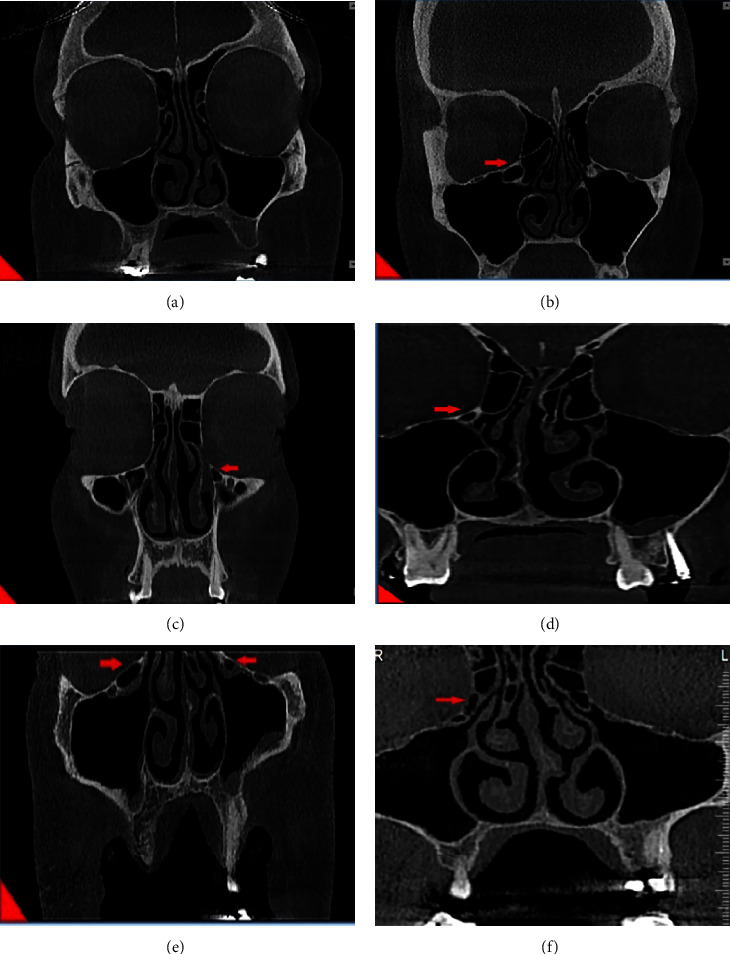
            A meticulous criteria for defining Haller cells as air cells was used, for any size located along medial portion of the orbital floor and/or lamina papyracea inferior to the bulla ethmoidalis and continuous with ethmoid capsula. The continuity with ethmoid capsula distinguishes Haller cells from infraorbital recess of maxillary sinus. Haller cell size was measured by its maximum mediolateral dimension. Maxillary ostium size is the distance between the most medial part of Haller cell and the uncinate process. Ostium and Haller cells based on size, are divided into 3 groups: small (less than 2 mm), medium (2–4 mm), and large (more than 4 mm). Infraorbital dehiscence is defined as the loss of bone density, and when the difference between dehiscence and thin bone wall is not recognizable, dehiscence diagnosis is acceptable (Figure 1).

            All collected data were executed by using the SPSS software (version 19), and the association between them was tested by the chi-squared test and Cochran–Mantel–Haenszel test.

            3. Results

            In this study, 120 patients (42.5% male and 57.5% female) were included from ages 18 to 79 with the average of 38.84. There was no significant relation between Haller cells and gender (p=0.682). Sixty-eight patients had Haller cells in their CBCTs, so the prevalence of Haller cells in this population was 56.7%, which included 64.7% unilateral and 35.3% bilateral. Both men and women were the same in the number of unilateral Haller cells, but in men, unilateral Haller cells were three times more than bilateral ones. Also, bilateral cases were twice in females. There was no significant relation between unilateral/bilateral and gender/age (p=0.186/p=0.419). Haller cells were categorized into three groups according to mediolateral dimensions: (a) small: less than 2 mm, (b) medium: 2–4 mm, and (c) large: more than 4 mm. Twenty percent of Haller cells were small, 70.67% were medium, and 9.33% were large, so the most prevalent size of Haller cells was medium (p ≤ 0.001). Even though the prevalence of Haller cells is various in different ages, we can assume all ages the same (p=0.282). Furthermore, there is no relation between size of Haller cells and gender/age (p=0.414/p=0.668).

            From 68 patients having Haller cells, 11 cases had orbital floor dehiscence. Fifty-two participants did not have Haller cells, also did not have orbital floor dehiscence. So, in this study, coexistence of Haller cells and orbital floor dehiscence is confirmed.

            4. Discussion

            This study estimated the prevalence of Haller cells in CBCT images almost high and about 56.7%. In some studies, an extremely variable range (2%–70.3%) has been reported for the prevalence of Haller cells [8, 1125]. Mathew et al. [6] reported a 60% prevalence for Haller cells and Khojastepour et al. [26] reported 68%, which are close to our study. Alkire and Bhattacharyya [13] also reported a 70.3% prevalence for Haller cells. This variability can be due to variation in subjects' race, age, sample size, observer's judgement regarding the presence of Haller cells in images, and different definitions for Haller cells in different studies. On the other hand, imaging technique also changes the results. Due to CBCT being a volumetric imaging technique, all the Haller cells in any size get captured; on the contrary, in multislice CT scans, small Haller cells could easily be missed in the interslice intervals [6]. The high percentage of Haller cells in this study can represent the high sensitivity of CBCT scan in the detection of small delicate bony structures.

            In this study, Haller cells were present unilaterally with statistical significance (64.7%). This finding is compatible with a large number of the previous studies [25, 7, 924, 2639].

            In Mathew's study, Haller cells were mostly present bilaterally, which was not statistically significant. The difference in the results can be due to the difference in the population and smaller sample size in Mathew's study (n = 50) than the present study (n = 120). Our study showed that the prevalence of Haller cell in women is slightly higher than in men. However, this difference is not statistically significant. Ozcan et al. also indicated that the prevalence of Haller cell is three times higher in female than in male [25]. These findings are consistent with the results of Khojastepour et al. [26], Raina et al. [1], and Basic et al.'s [40] studies.

            The most prevalent Haller cells observed in our study were medium sized (2–4 mm) with a significant difference. In the study of Dhillon and Kalra [32], about 51% of cases were large (>4 mm). The difference between these two studies can be justified by the variation in the ethnic characteristics of the populations studied and the sample size.

            In this study, more than 55% of positive cases were under 40 years old. However, the difference in the distribution of Haller cell prevalence by age was not statistically significant. In Raina et al.'s study, 64.6% of Haller cells were observed in subjects from ages 18 to 30, which is consistent with our study results [1].

            Various studies have assessed the relationship between presence of Haller cell and maxillary sinus drainage and pathologies such as sinusitis [4144]. Although presence of Haller cells may interfere with normal sinus drainage, Suzuki-Yamazaki performed a successful sinus lift procedure in a patient with large Haller cell [41]. To the best of our knowledge, this is one of the first studies assessing the relationship of Haller cell size and orbital floor dehiscence in Iranian population. This study confirmed the existence of a significant relationship between the presence of Haller cells and dehiscence of orbital floor (p=0.002). Lack of bone density and presence of only a mucoperiosteal lining that separates Haller cell from orbital space were considered as dehiscence. Diagnosis of this issue in CBCT images is really important because the presence of orbital floor dehiscence predisposes orbital space to Haller cell diseases or makes it vulnerable in osteomeatal complex surgery.

            Seberchets et al. published an article in 2000 containing three case reports of unilateral orbital cellulitis caused by inflammation of the Haller cells [45]. They declared that any pathologic lesion related to Haller cells should be considered as a potential for unilateral eye cellulitis. Given that there is no lymphatic drainage system in the eye, they propounded a hypothesis that infection spreads through orbital floor dehiscence, lamina papyracea, or sutures of the medial part of orbital floor. Mathew et al.'s study also showed a significant relationship between Haller cells and orbital floor dehiscence; both of these results support our study (3).

            So according to this study, we suggest that anytime an inflamed Haller cell is observed in CBCT. Simultaneous presence of orbital floor dehiscence should be expected.

            5. Conclusion

            This study estimated the prevalence of Haller cells in CBCT high (56.7%) and noticeable and also showed that statistically, there is a significant relationship between Haller cells and orbital floor dehiscence. It can be concluded that CBCT can be a useful imaging modality for evaluating the anatomical aspect of sinonasal bone complex due to its high accuracy and lower radiation dose.

            6. Limitations and Suggestions

            6.1. Limitation

            Due to the nature of this study (in vitro) and method of collecting the images (CBCTs stored in university's archive), there was no ethical limitation.

            6.2. Recommendations

            For increasing the accuracy and efficiency of the study, we suggest a larger population with more samples available.

            Different resolutions should be used in CBCT imaging for better diagnosis of Haller cells and orbital floor dehiscence.

            Data Availability

            The datasets used and/or analyzed during the current study are available from the corresponding author upon request.

            Ethical Approval

            The study protocol was approved by the Institutional Review Board of Shahid Beheshti University of Medical Sciences.

            Consent

            All authors gave their consent for the publication of this manuscript.

            Disclosure

            A preprint has previously been published [46].

            Conflicts of Interest

            The authors declare that they have no conflicts of interest.

            Authors' Contributions

            Mahkameh Moshfeghi designed and supervised the study process. Hamidreza Dehini performed data acquisition and investigation. Ghazizaeh Ahsaie M participated in the data analysis and interpretation. Ghazizadeh Ahsaie drafted the manuscript. All the authors have reviewed and approved the final manuscript.

            References

            1. Raina A., Guledgud M. V., Patil K.. Infraorbital ethmoid (Haller’s) cells: a panoramic radiographic study. Dentomaxillofacial Radiology . 2012. Vol. 41(4):305–308. [Cross Ref] [2-s2.0-84860115770] [PubMed]

            2. Ahmad M., Khurana N., Jaberi J., Sampair C., Kuba R. K.. Prevalence of infraorbital ethmoid (Haller’s) cells on panoramic radiographs. Oral Surgery, Oral Medicine, Oral Pathology, Oral Radiology and Endodontics . 2006. Vol. 101(5):658–661. [Cross Ref] [2-s2.0-33646053472] [PubMed]

            3. Kantarci M., Karasen R., Alper F., Onbas O., Okur A., Karaman A.. Remarkable anatomic variations in paranasal sinus region and their clinical importance. European Journal of Radiology . 2004. Vol. 50(3):296–302. [Cross Ref] [2-s2.0-2442540535] [PubMed]

            4. Lerdlum S., Vachiranubhap B.. Prevalence of anatomic variation demonstrated on screening sinus computed tomography and clinical correlation. Journal of the Medical Association of Thailand= Chotmaihet thangphaet . 2005. Vol. 88(4):S110–S115

            5. Leunig A.. Anatomic variations of the sinuses; multiplanar CT-analysis in 641 patients. Laryngo-Rhino-Otologie . 2008. Vol. 87(7):482–489. [Cross Ref] [2-s2.0-49749152085] [PubMed]

            6. Mathew R., Omami G., Hand A., Fellows D., Lurie A.. Cone beam CT analysis of Haller cells: prevalence and clinical significance. Dentomaxillofacial Radiology . 2013. Vol. 42(9)[20130055] [Cross Ref] [2-s2.0-84885602042]

            7. Kayalioglu G., Oyar O., Govsa F.. Nasal cavity and paranasal sinus bony variations: a computed tomographic study. Rhinology . 2000. Vol. 38(3):108–113. [PubMed]

            8. Hui L., Hung K. F., Yeung A. W. K., von Arx T., Leung Y. Y., Bornstein M. M.. Anatomical variations of the ethmoid sinuses and their association with health or pathology of the ethmoid and maxillary sinuses in a Southern Chinese population: an analysis using cone-beam computed tomography. Imaging science in dentistry . 2022. Vol. 52(1):p. 109[Cross Ref]

            9. Khayam E.. The prevalence of ethmoidal infraorbital cells in panoramic radiography. American Journal of Research Communication . 2013. Vol. 1:109–118

            10. Valizadeh S.. Visibility of infraorbital ethmoid air cell on panoramic radiographs. Shahid Beheshti University Dental Journal . 2010. Vol. 28(1):7–11

            11. Fadda G. L., Rosso S., Aversa S., Petrelli A., Ondolo C., Succo G.. Multiparametric statistical correlations between paranasal sinus anatomic variations and chronic rhinosinusitis. Acta Otorhinolaryngologica Italica: organo ufficiale della Societa italiana di otorinolaringologia e chirurgia cervico-facciale . 2012. Vol. 32(4):244–251. [PubMed]

            12. Mamatha H.. Variations of ostiomeatal complex and its applied anatomy: a CT scan study. Indian Journal of Science and Technology . 2010. Vol. 3(8):904–907. [Cross Ref]

            13. Alkire B. C., Bhattacharyya N.. An assessment of sinonasal anatomic variants potentially associated with recurrent acute rhinosinusitis. The Laryngoscope . 2010. Vol. 120(3):631–634. [Cross Ref] [2-s2.0-77949526930] [PubMed]

            14. Miranda C. M. N. R. D., Padilha I. G.. Anatomical variations of paranasal sinuses at multislice computed tomography: what to look for. Radiologia Brasileira . 2011. Vol. 44(4):256–262

            15. Lloyd G. A. S.. CT of the paranasal sinuses: study of a control series in relation to endoscopic sinus surgery. Journal of Laryngology and Otology . 1990. Vol. 104(6):477–481. [Cross Ref] [2-s2.0-0025294705] [PubMed]

            16. Lloyd G. A. S., Lund V. J., Scadding G. K.. CT of the paranasal sinuses and functional endoscopic surgery: a critical analysis of 100 symptomatic patients. Journal of Laryngology and Otology . 1991. Vol. 105(3):181–185. [Cross Ref] [2-s2.0-0025827955] [PubMed]

            17. Scribano E., Ascenti G., Cascio F., Racchiusa S., Salamone I.. Computerized tomography in the evaluation of anatomic variations of the ostiomeatal complex. La Radiologia medica . 1993. Vol. 86(3):195–199. [PubMed]

            18. Yousem D. M.. Imaging of sinonasal inflammatory disease. Radiology . 1993. Vol. 188(2):303–314. [Cross Ref] [2-s2.0-0027255055] [PubMed]

            19. Wanamaker H. H.. Role of Haller’s cell in headache and sinus disease: a case report. Otolaryngology-Head and Neck Surgery . 1996. Vol. 114(2):324–327. [Cross Ref] [2-s2.0-0030041392] [PubMed]

            20. Tonai A., Baba S.. Anatomic variations of the bone in sinonasal CT. Acta oto-laryngologica. Supplementum . 1996. Vol. 525:9–13. [PubMed]

            21. Stackpole S. A., Edelstein D. R.. The anatomic relevance of the Haller cell in sinusitis. American Journal of Rhinology . 1997. Vol. 11(3):219–224. [Cross Ref] [2-s2.0-0031135998] [PubMed]

            22. Perez-Pinas I., Sabate J., Carmona A., Catalina-Herrera C. J., Jimenez-Castellanos J.. Anatomical variations in the human paranasal sinus region studied by CT. Journal of Anatomy . 2000. Vol. 197(2):221–227. [Cross Ref] [2-s2.0-0033857420] [PubMed]

            23. Zinreich S.. The ostiomeatal complex and functional endoscopic surgery. Head and Neck Imaging . 2003. 4. Missouri, MO, USA: Mosby Elsevier. p. 149–173

            24. Wani A. A., Kanotra S., Lateef M., Ahmad R., Qazi S. M., Ahmad S.. CT scan evaluation of the anatomical variations of the ostiomeatal complex. Indian Journal of Otolaryngology and Head and Neck Surgery . 2009. Vol. 61(3):163–168. [Cross Ref] [2-s2.0-70350147421] [PubMed]

            25. Özcan İ., Goksel S., Cakir-Karabas H., Unsal G.. CBCT analysis of haller cells: relationship with accessory maxillary ostium and maxillary sinus pathologies. Oral Radiology . 2021. Vol. 37(3):502–506. [Cross Ref] [PubMed]

            26. Khojastepour L., Mirhadi S., Mesbahi S. A.. Anatomical variations of ostiomeatal complex in CBCT of patients seeking rhinoplasty. Journal of Dentistry . 2015. Vol. 16(1):42–48. [PubMed]

            27. Mazza D., Bontempi E., Guerrisi A., et al.. Paranasal sinuses anatomic variants: 64-slice CT evaluation. Minerva Stomatologica . 2007. Vol. 56(6):311–318. [PubMed]

            28. Jaju P. P., Jaju S. P.. Clinical utility of dental cone-beam computed tomography: current perspectives. Clinical, Cosmetic and Investigational Dentistry . 2014. Vol. 6:29–43. [Cross Ref] [2-s2.0-84898041261] [PubMed]

            29. White S. C., Pharoah M. J.. Oral Radiology-E-Book: Principles and Interpretation . 2014. Amsterdam, Netherlands: Elsevier Health Sciences.

            30. Schwarz M. S., Rothman S. L., Rhodes M. L., Chafetz N.. Computed tomography: part I. preoperative assessment of the mandible for endosseous implant surgery. The International Journal of Oral and Maxillofacial Implants . 1987. Vol. 2(3):137–141. [PubMed]

            31. Timock A. M., Cook V., McDonald T., et al.. Accuracy and reliability of buccal bone height and thickness measurements from cone-beam computed tomography imaging. American Journal of Orthodontics and Dentofacial Orthopedics . 2011. Vol. 140(5):734–744. [Cross Ref] [2-s2.0-80155126201] [PubMed]

            32. Dhillon J. K., Kalra G.. Cone beam computed tomography: an innovative tool in pediatric dentistry. Journal of Pediatric Dentistry . 2013. Vol. 1(2):p. 27[Cross Ref]

            33. Damstra J., Fourie Z., Huddleston Slater J. J., Ren Y.. Accuracy of linear measurements from cone-beam computed tomography-derived surface models of different voxel sizes. American Journal of Orthodontics and Dentofacial Orthopedics . 2010. Vol. 137(1):16. e1–e16.e6. [Cross Ref] [2-s2.0-72649103006]

            34. Özen T., Kamburoglu K., Cebeci A. R. I., Yuksel S. P., Paksoy C. S.. Interpretation of chemically created periapical lesions using 2 different dental cone-beam computerized tomography units, an intraoral digital sensor, and conventional film. Oral Surgery, Oral Medicine, Oral Pathology, Oral Radiology and Endodontics . 2009. Vol. 107(3):426–432. [Cross Ref] [2-s2.0-59849113724] [PubMed]

            35. Oz U., Orhan K., Abe N.. Comparison of linear and angular measurements using two-dimensional conventional methods and three-dimensional cone beam CT images reconstructed from a volumetric rendering program in vivo. Dentomaxillofacial Radiology . 2011. Vol. 40(8):492–500. [Cross Ref] [2-s2.0-82755170427] [PubMed]

            36. Kamburoğlu K., Kolsuz E., Kurt H., Kilic C., Ozen T., Semra Paksoy C.. Accuracy of CBCT measurements of a human skull. Journal of Digital Imaging . 2011. Vol. 24(5):787–793. [Cross Ref] [2-s2.0-84855592424] [PubMed]

            37. Tomasi C., Bressan E., Corazza B., Mazzoleni S., Stellini E., Lith A.. Reliability and reproducibility of linear mandible measurements with the use of a cone-beam computed tomography and two object inclinations. Dentomaxillofacial Radiology . 2011. Vol. 40(4):244–250. [Cross Ref] [2-s2.0-79955040862] [PubMed]

            38. Sheikhi M., Ghorbanizadeh S., Abdinian M., Goroohi H., Badrian H.. Accuracy of linear measurements of galileos cone beam computed tomography in normal and different head positions. International journal of dentistry . 2012. Vol. 2012:6[214954] [Cross Ref] [2-s2.0-84864953120]

            39. Earwaker J.. Anatomic variants in sinonasal CT. RadioGraphics . 1993. Vol. 13(2):381–415. [Cross Ref] [2-s2.0-0027566287] [PubMed]

            40. Bašić N., Basic V., Jukic T., Basic M., Jelic M., Hat J.. Computed tomographic imaging to determine the frequency of anatomical variations in pneumatization of the ethmoid bone. European Archives of Oto-Rhino-Laryngology . 1999. Vol. 256(2):69–71. [Cross Ref] [2-s2.0-0033003087] [PubMed]

            41. Suzuki-Yamazaki M.. A successful treatment regimen for the prevention of sinusitis after maxillary sinus floor elevation surgery in a high-risk case. Case Reports in Otolaryngology . 2020. Vol. 2020:3[6869805] [Cross Ref]

            42. Şahin B., Comoglu S., Sonmez S., Deger K., Keles Turel M. N.. Paranasal sinus fungus ball, anatomical variations and dental pathologies: is there any relation? Turkish Archives of Otolaryngology . 2022. Vol. 60(1):23–28. [Cross Ref]

            43. Srinivas C. V., Kauser S.. Anatomy and variations of onodi cells and haller cells: a hrct cum clinical analysis in sinonasal disease and polyposis. Indian Journal of Otolaryngology and Head and Neck Surgery . 2021. Vol. 74(S2):1683–1689. [Cross Ref] [PubMed]

            44. Papadopoulou A.-M., Chrysikos D., Samolis A., Tsakotos G., Troupis T.. Anatomical variations of the nasal cavities and paranasal sinuses: a systematic review. Cureus . 2021. Vol. 13(1)[e12727] [Cross Ref]

            45. Sebrechts H., Vlaminck S., Casselman J.. Orbital edema resulting from Haller’s cell pathology: 3 case reports and review of literature. Acta Oto-Rhino-Laryngologica Belgica . 2000. Vol. 54(1):39–43. [PubMed]

            46. Moshfeghi M., Ahsaie M. G.. Cone Beam CT Analysis of Haller Cells: Prevalence and Relationship with Orbital Floor Dehiscence . 2021

            Floating objects

            Figure 1

            Detection of Haller cell in coronal view CBCT. (a) No Haller cell is detected. (b) Large Haller cell on the right side. (c) Medium sized Haller cell on the left. (d) Small-sized Haller cell on the right. (e) Bilateral presence of Haller cell with two different sizes. (f) Dehiscence of inferomedial border of orbit due to the presence.

            Author and article information

            Contributors
            Journal
            Int J Dent
            Int J Dent
            ijd
            International Journal of Dentistry
            Hindawi
            1687-8728
            1687-8736
            2023
            31 January 2023
            : 2023
            : 5200152
            Affiliations
            1Department of Oral and Maxillofacial Radiology, School of Dentistry, Shahid Beheshti University of Medical Sciences, Tehran, Iran
            2School of Dentistry, Shahid Beheshti University of Medical Sciences, Tehran, Iran
            Author notes

            Academic Editor: Mario Dioguardi

            Author information
            https://orcid.org/0000-0002-1997-9437
            https://orcid.org/0000-0002-1107-6487
            https://orcid.org/0000-0001-7028-748X
            Article
            10.1155/2023/5200152
            9904925
            72c97ceb-b734-4855-b44b-ae9f10a0eb58
            Copyright © 2023 Mahkameh Moshfeghi et al.

            This is an open access article distributed under the Creative Commons Attribution License, which permits unrestricted use, distribution, and reproduction in any medium, provided the original work is properly cited.

            History
            : 30 March 2022
            : 26 July 2022
            : 18 January 2023
            Categories
            Research Article

            Dentistry

            Comments

            Comment on this article