59
views
0
recommends
+1 Recommend
0 collections
    0
    shares
      • Record: found
      • Abstract: found
      • Article: found
      Is Open Access

      Cone Beam CT Analysis of Haller Cells: Prevalence and Relationship with Orbital Floor Dehiscence

      research-article
      1 , 2 , 1 ,
      International Journal of Dentistry
      Hindawi

      Read this article at

      Bookmark
          There is no author summary for this article yet. Authors can add summaries to their articles on ScienceOpen to make them more accessible to a non-specialist audience.

          Abstract

          Materials and Methods

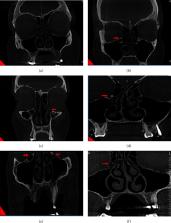
          CBCT images of 120 patients were interpreted in coronal plane for the presence of Haller cells and orbital floor dehiscence. The prevalence of Haller cell, presence of dehiscence, unilateral, or bilateral frequency were assessed. In addition, the size was categorized in three groups of small, medium, and large. Chi-square and Cochran–Mantel–Haenszel tests were used for statistical analysis of the data, and p < 0.05 was considered to be significant.

          Results

          A total of 51 male and 69 female with mean ± SD age of 38.84 ± 68.14 were assessed. The overall prevalence of Haller cells was 56.7%, of which 44 (64.7%) were unilateral and 24 were bilateral (35.3%). The majority of the cells (70.7%) were seen in medium (2–4 mm) sized. There was a significant association between Haller cells and orbital floor dehiscence ( p ≤ 0.001).

          Conclusion

          The prevalence of Haller cells was remarkably high and the presence of Haller cells was strongly associated with ipsilateral orbital floor dehiscence. Based on the findings of this study, CBCT can be useful in delineation of the bony anatomy of sinonasal complex.

          Related collections

          Most cited references46

          • Record: found
          • Abstract: found
          • Article: not found

          Accuracy and reliability of buccal bone height and thickness measurements from cone-beam computed tomography imaging.

          Cone-beam computed tomography (CBCT) imaging has broadened opportunities for examining morphologic aspects of the craniofacial complex, including alveolar bone, but limitations of the technology have yet to be defined. Through the use of comparisons with direct measurements, the purpose of this study was to investigate the accuracy and reliability of buccal alveolar bone height and thickness measurements derived from CBCT images. Twelve embalmed cadaver heads (5 female, 7 male; mean age: 77 years) were scanned with an i-CAT 17-19 unit (Imaging Sciences International, Hatfield, Pa) at 0.3 mm voxel size. Buccal alveolar bone height and thickness measurements of 65 teeth were made in standardized radiographic slices and compared with direct measurements made by dissection. All measurements were repeated 3 times by 2 independent raters and examined for intrarater and interrater reliability. Measurement means were compared with 2-tailed t tests. Agreement between direct and CBCT measurements was assessed by concordance correlation coefficients, Pearson correlation coefficients, and Bland-Altman plots. Intrarater reliability was high as were interrater correlations for all measurements (≥0.97) except CBCT buccal bone thickness (0.90). CBCT measurements did not differ significantly from direct measurements, and there was no pattern of underestimation or overestimation. The mean absolute differences were 0.30 mm in buccal bone height and 0.13 mm in buccal bone thickness with 95% limits of agreement of -0.77 to 0.81 mm, and -0.32 to 0.38 mm, respectively. Agreement between the 2 methods was higher for the measurements of buccal bone height than buccal bone thickness, as demonstrated by concordance correlation coefficients of 0.98 and 0.86, respectively. For the protocol used in this study, CBCT can be used to quantitatively assess buccal bone height and buccal bone thickness with high precision and accuracy. Comparing the 2 sets of CBCT measurements, buccal bone height had greater reliability and agreement with direct measurements than did the buccal bone thickness measurements. Copyright © 2011 American Association of Orthodontists. Published by Mosby, Inc. All rights reserved.
            Bookmark
            • Record: found
            • Abstract: found
            • Article: not found

            Anatomic variants in sinonasal CT.

            W Earwaker (1993)
            The computed tomographic scans obtained in a series of 800 patients referred for evaluation for functional endoscopic sinus surgery were examined to determine the prevalence and significance of anatomic variants. Fifty-two normal variants were identified within two major groupings of primary bony abnormalities and sinus air cell extensions. Although 743 (93%) patients had one or more variants, the nature of the variants in many instances was such that 325 (41%) patients could be considered "endoscopically" normal. Among the remaining cases, variations of the septum and middle turbinates, with or without anterior ethmoid sinus extensions, were found, usually in recognizable combinations, that could produce significant obstruction of the drainage pathways. However, where such obstructive patterns existed, an equal prevalence of patients with and without sinus disease was found in the presence of the same variant combination. Thus, the presence of anatomic variations, singly or in combination, does not represent a disease state per se.
              Bookmark
              • Record: found
              • Abstract: found
              • Article: not found

              Remarkable anatomic variations in paranasal sinus region and their clinical importance.

              With the advent of functional endoscopic sinus surgery (FESS) and coronal computed tomography (CT) imaging, considerable attention has been directed toward paranasal region anatomy. Detailed knowledge of anatomic variations in paranasal sinus region is critical for surgeons performing endoscopic sinus surgery as well as for the radiologist involved in the preoperative work-up. To be in the known anatomical variants with some accompanying pathologies, directly influence the success of diagnostic and therapeutic management of paranasal sinus diseases. A review of 512 (1024 sides) paranasal sinus tomographic scans was carried out to expose remarkable anatomic variations of this region. We used only coronal sections, but for some cases to clear exact diagnosis, additional axial CT scan, magnetic resonance imaging (MRI) and nasal endoscopy were also performed. In this pictorial essay, rates of remarkable anatomic variations in paranasal region were displayed. The images of some interesting cases were illustrated, such as the Onodi cell in which isolated mucocele caused loss of visual acuity, agger nasi cell, Haller's cell, uncinate bulla, giant superior concha bullosa, inferior concha bullosa, bilateral carotid artery protrusion into sphenoid sinus, maxillary sinus agenesis, bilateral secondary middle turbinate (SMT) and sphenomaxillary plate. The clinical importance of all these variations were discussed under the light of the literature. It was suggested that remarkable anatomic variations of paranasal region and their possible pathologic consequences should be well defined in order to improve success of management strategies, and to avoid potential complications of endoscopic sinus surgery. The radiologist must pay close attention to anatomical variations in the preoperative evaluation.
                Bookmark

                Author and article information

                Contributors
                Journal
                Int J Dent
                Int J Dent
                ijd
                International Journal of Dentistry
                Hindawi
                1687-8728
                1687-8736
                2023
                31 January 2023
                : 2023
                : 5200152
                Affiliations
                1Department of Oral and Maxillofacial Radiology, School of Dentistry, Shahid Beheshti University of Medical Sciences, Tehran, Iran
                2School of Dentistry, Shahid Beheshti University of Medical Sciences, Tehran, Iran
                Author notes

                Academic Editor: Mario Dioguardi

                Author information
                https://orcid.org/0000-0002-1997-9437
                https://orcid.org/0000-0002-1107-6487
                https://orcid.org/0000-0001-7028-748X
                Article
                10.1155/2023/5200152
                9904925
                72c97ceb-b734-4855-b44b-ae9f10a0eb58
                Copyright © 2023 Mahkameh Moshfeghi et al.

                This is an open access article distributed under the Creative Commons Attribution License, which permits unrestricted use, distribution, and reproduction in any medium, provided the original work is properly cited.

                History
                : 30 March 2022
                : 26 July 2022
                : 18 January 2023
                Categories
                Research Article

                Dentistry
                Dentistry

                Comments

                Comment on this article