43
views
0
recommends
+1 Recommend
0 collections
    0
    shares
      • Record: found
      • Abstract: found
      • Article: found
      Is Open Access

      The huntingtin N17 domain is a multifunctional CRM1 and Ran-dependent nuclear and cilial export signal

      research-article
      , , , *
      Human Molecular Genetics
      Oxford University Press

      Read this article at

      Bookmark
          There is no author summary for this article yet. Authors can add summaries to their articles on ScienceOpen to make them more accessible to a non-specialist audience.

          Abstract

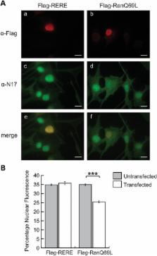
          The first 17 amino acids of Huntington’s disease (HD) protein, huntingtin, comprise an amphipathic alpha-helical domain that can target huntingtin to the endoplasmic reticulum (ER). N17 is phosphorylated at two serines, shown to be important for disease development in genetic mouse models, and shown to be modified by agents that reverse the disease phenotype in an HD mouse model. Here, we show that the hydrophobic face of N17 comprises a consensus CRM1/exportin-dependent nuclear export signal, and that this nuclear export activity can be affected by serine phospho-mimetic mutants. We define the precise residues that comprise this nuclear export sequence (NES) as well as the interaction of the NES, but not phospho-mimetic mutants, with the CRM1 nuclear export factor. We show that the nuclear localization of huntingtin depends upon the RanGTP/GDP gradient, and that N17 phosphorylation can also distinguish localization of endogenous huntingtin between the basal body and stalk of the primary cilium. We present a mechanism and multifunctional role for N17 in which phosphorylation of N17 not only releases huntingtin from the ER to allow nuclear entry, but also prevents nuclear export during a transient stress response event to increase the levels of nuclear huntingtin and to regulate huntingtin access to the primary cilium. Thus, N17 is a master localization signal of huntingtin that can mediate huntingtin localization between the cytoplasm, nucleus and primary cilium. This localization can be regulated by signaling, and is misregulated in HD.

          Related collections

          Most cited references49

          • Record: found
          • Abstract: found
          • Article: not found

          Full-length human mutant huntingtin with a stable polyglutamine repeat can elicit progressive and selective neuropathogenesis in BACHD mice.

          To elucidate the pathogenic mechanisms in Huntington's disease (HD) elicited by expression of full-length human mutant huntingtin (fl-mhtt), a bacterial artificial chromosome (BAC)-mediated transgenic mouse model (BACHD) was developed expressing fl-mhtt with 97 glutamine repeats under the control of endogenous htt regulatory machinery on the BAC. BACHD mice exhibit progressive motor deficits, neuronal synaptic dysfunction, and late-onset selective neuropathology, which includes significant cortical and striatal atrophy and striatal dark neuron degeneration. Power analyses reveal the robustness of the behavioral and neuropathological phenotypes, suggesting BACHD as a suitable fl-mhtt mouse model for preclinical studies. Additional analyses of BACHD mice provide novel insights into how mhtt may elicit neuropathogenesis. First, unlike previous fl-mhtt mouse models, BACHD mice reveal that the slowly progressive and selective pathogenic process in HD mouse brains can occur without early and diffuse nuclear accumulation of aggregated mhtt (i.e., as detected by immunostaining with the EM48 antibody). Instead, a relatively steady-state level of predominantly full-length mhtt and a small amount of mhtt N-terminal fragments are sufficient to elicit the disease process. Second, the polyglutamine repeat within fl-mhtt in BACHD mice is encoded by a mixed CAA-CAG repeat, which is stable in both the germline and somatic tissues including the cortex and striatum at the onset of neuropathology. Therefore, our results suggest that somatic repeat instability does not play a necessary role in selective neuropathogenesis in BACHD mice. In summary, the BACHD model constitutes a novel and robust in vivo paradigm for the investigation of HD pathogenesis and treatment.
            Bookmark
            • Record: found
            • Abstract: found
            • Article: not found

            Dominant phenotypes produced by the HD mutation in STHdh(Q111) striatal cells.

            Lengthening a glutamine tract in huntingtin confers a dominant attribute that initiates degeneration of striatal neurons in Huntington's disease (HD). To identify pathways that are candidates for the mutant protein's abnormal function, we compared striatal cell lines established from wild-type and Hdh(Q111) knock-in embryos. Alternate versions of full-length huntingtin, distinguished by epitope accessibility, were localized to different sets of nuclear and perinuclear organelles involved in RNA biogenesis and membrane trafficking. However, mutant STHdh(Q111) cells also exhibited additional forms of the full-length mutant protein and displayed dominant phenotypes that did not mirror phenotypes caused by either huntingtin deficiency or excess. These phenotypes indicate a disruption of striatal cell homeostasis by the mutant protein, via a mechanism that is separate from its normal activity. They also support specific stress pathways, including elevated p53, endoplasmic reticulum stress response and hypoxia, as potential players in HD.
              Bookmark
              • Record: found
              • Abstract: found
              • Article: not found

              Huntingtin acts in the nucleus to induce apoptosis but death does not correlate with the formation of intranuclear inclusions.

              The mechanisms by which mutant huntingtin induces neurodegeneration were investigated using a cellular model that recapitulates features of neurodegeneration seen in Huntington's disease. When transfected into cultured striatal neurons, mutant huntingtin induces neurodegeneration by an apoptotic mechanism. Antiapoptotic compounds or neurotrophic factors protected neurons against mutant huntingtin. Blocking nuclear localization of mutant huntingtin suppressed its ability to form intranuclear inclusions and to induce neurodegeneration. However, the presence of inclusions did not correlate with huntingtin-induced death. The exposure of mutant huntingtin-transfected striatal neurons to conditions that suppress the formation of inclusions resulted in an increase in mutant huntingtin-induced death. These findings suggest that mutant huntingtin acts within the nucleus to induce neurodegeneration. However, intranuclear inclusions may reflect a cellular mechanism to protect against huntingtin-induced cell death.
                Bookmark

                Author and article information

                Journal
                Hum Mol Genet
                Hum. Mol. Genet
                hmg
                hmg
                Human Molecular Genetics
                Oxford University Press
                0964-6906
                1460-2083
                1 April 2013
                7 January 2013
                7 January 2013
                : 22
                : 7
                : 1383-1394
                Affiliations
                Department of Biochemistry and Biomedical Sciences, McMaster University , 1200 Main Street West, Hamilton, ON, CanadaL8N3Z5
                Author notes
                [* ]To whom correspondence should be addressed. Tel: +905-525-9140; Fax: +905-522-9033; Email: truantr@ 123456mcmaster.ca
                Article
                dds554
                10.1093/hmg/dds554
                3596850
                23297360
                fffc39b7-9874-46a0-9264-8583e68274a7
                © The Author 2013. Published by Oxford University Press.

                This is an Open Access article distributed under the terms of the Creative Commons Attribution License ( http://creativecommons.org/licenses/by-nc/3.0/), which permits non-commercial use, distribution, and reproduction in any medium, provided the original work is properly cited. For commercial re-use, please contact journals.permission@oup.com.

                History
                : 16 October 2012
                : 14 December 2012
                : 27 December 2012
                Categories
                Articles

                Genetics
                Genetics

                Comments

                Comment on this article