0
views
0
recommends
+1 Recommend
0 collections
    0
    shares
      • Record: found
      • Abstract: found
      • Article: found
      Is Open Access

      Establishment of a bi-layered tissue engineered conjunctiva using a 3D-printed melt electrowritten poly-(ε-caprolactone) scaffold

      research-article

      Read this article at

      Bookmark
          There is no author summary for this article yet. Authors can add summaries to their articles on ScienceOpen to make them more accessible to a non-specialist audience.

          Abstract

          Purpose

          To utilize melt electrowriting (MEW) technology using poly-(ε-caprolactone) (PCL) coupled with a 2-step co-culturing strategy for the development of a conjunctival bi-layer synthetic construct.

          Methods

          Melt electrowritten scaffolds using PCL were fabricated using an in-house-built MEW printer. Human conjunctival stromal cells (CjSCs) and epithelial cells (CjECs) were isolated from donor tissue. A 2-step co-culture method was done by first seeding the CjSCs and culturing for 4 weeks to establish a stromal layer, followed by CjECs and co-culturing for 2 more weeks. Cultured cells were each characterized by morphology and marker expression on immunofluorescence and qPCR. The produced construct was assessed for cellular proliferation using viability assays. The bi-layer morphology was assessed using scanning electron microscopy (SEM), confocal microscopy, and immunofluorescence imaging. The expression of extracellular matrix components and TGF-b was evaluated using qPCR.

          Results

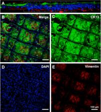
          CjSCs were spindle-shaped and vimentin + while CjECs were polygonal and CK13 + . CjSCs showed consistent proliferation and optimal adherence with the scaffold at the 4-week culture mark. A 2-layered construct consisting of a CjSC-composed stromal layer and a CjEC-composed epithelial layer was appreciated on confocal microscopy, SEM, and immunofluorescence. CjSCs secreted collagens (types I, V, VI) but at differing amounts from natural tissue while TGF-b production was comparable.

          Conclusion

          The 3D-printed melt electrowritten PCL scaffold paired with the 2-step co-culturing conditions of the scaffold allowed for the first approximation of a bi-layered stromal and epithelial reconstruction of the conjunctiva that can potentially improve the therapeutic arsenal in ocular surface reconstruction.

          Supplementary Information

          The online version contains supplementary material available at 10.1007/s10792-022-02418-y.

          Related collections

          Most cited references43

          • Record: found
          • Abstract: not found
          • Article: not found

          The return of a forgotten polymer—Polycaprolactone in the 21st century

            Bookmark
            • Record: found
            • Abstract: found
            • Article: not found

            Direct writing by way of melt electrospinning.

            Melt electrospun fibers of poly(ϵ-caprolactone) are accurately deposited using an automated stage as the collector. Matching the translation speed of the collector to the speed of the melt electrospinning jet establishes control over the location of fiber deposition. In this sense, melt electrospinning writing can be seen to bridge the gap between solution electrospinning and direct writing additive manufacturing processes. Copyright © 2011 WILEY-VCH Verlag GmbH & Co. KGaA, Weinheim.
              Bookmark
              • Record: found
              • Abstract: found
              • Article: not found

              Electrospun poly(epsilon-caprolactone) microfiber and multilayer nanofiber/microfiber scaffolds: characterization of scaffolds and measurement of cellular infiltration.

              The physical and spatial architectural geometries of electrospun scaffolds are important to their application in tissue engineering strategies. In this work, poly(epsilon-caprolactone) microfiber scaffolds with average fiber diameters ranging from 2 to 10 microm were individually electrospun to determine the parameters required for reproducibly fabricating scaffolds. As fiber diameter increased, the average pore size of the scaffolds, as measured by mercury porosimetry, increased (values ranging from 20 to 45 microm), while a constant porosity was observed. To capitalize on both the larger pore sizes of the microfiber layers and the nanoscale dimensions of the nanofiber layers, layered scaffolds were fabricated by sequential electrospinning. These scaffolds consisted of alternating layers of poly(epsilon-caprolactone) microfibers and poly(epsilon-caprolactone) nanofibers. By electrospinning the nanofiber layers for different lengths of time, the thickness of the nanofiber layers could be modulated. Bilayered constructs consisting of microfiber scaffolds with varying thicknesses of nanofibers on top were generated and evaluated for their potential to affect rat marrow stromal cell attachment, spreading, and infiltration. Cell attachment after 24 h did not increase with increasing number of nanofibers, but the presence of nanofibers enhanced cell spreading as evidenced by stronger F-actin staining. Additionally, increasing the thickness of the nanofiber layer reduced the infiltration of cells into the scaffolds under both static and flow perfusion culture for the specific conditions tested. The scaffold design presented in this study allows for cellular infiltration into the scaffolds while at the same time providing nanofibers as a physical mimicry of extracellular matrix.
                Bookmark

                Author and article information

                Contributors
                victor.perez.quinones@duke.edu
                Journal
                Int Ophthalmol
                Int Ophthalmol
                International Ophthalmology
                Springer Netherlands (Dordrecht )
                0165-5701
                1573-2630
                6 August 2022
                6 August 2022
                2023
                : 43
                : 1
                : 215-232
                Affiliations
                [1 ]GRID grid.419851.0, Department of Ophthalmology, , Ocular Surface Center, Bascom Palmer Eye Institute, University of Miami Miller School of Medicine, ; Miami, FL USA
                [2 ]GRID grid.26790.3a, ISNI 0000 0004 1936 8606, Interdisciplinary Stem Cell Institute, , University of Miami Miller School of Medicine, ; Miami, FL USA
                [3 ]GRID grid.412465.0, Department of Ophthalmology, , Second Affiliated Hospital, Zhejiang University School of Medicine, ; Hangzhou, China
                [4 ]GRID grid.38142.3c, ISNI 000000041936754X, Massachusetts Eye and Ear Infirmary, , Harvard Medical School, ; Boston, MA USA
                [5 ]GRID grid.26009.3d, ISNI 0000 0004 1936 7961, Department of Ophthalmology, , Foster Center for Ocular Immunology, Distinguished Stephen and Frances Foster Chair in Ocular Immunology, Duke Eye Center, ; 2351 Erwin Road, Durham, NC 27705 USA
                [6 ]GRID grid.1024.7, ISNI 0000000089150953, Institute for Health and Biomedical Innovation (IHBI), , Queensland University of Technology (QUT), ; Brisbane, QLD Australia
                Article
                2418
                10.1007/s10792-022-02418-y
                9902434
                35932420
                ffd413b1-28e1-4bfe-988f-dbbee639d42d
                © The Author(s) 2022

                Open AccessThis article is licensed under a Creative Commons Attribution 4.0 International License, which permits use, sharing, adaptation, distribution and reproduction in any medium or format, as long as you give appropriate credit to the original author(s) and the source, provide a link to the Creative Commons licence, and indicate if changes were made. The images or other third party material in this article are included in the article's Creative Commons licence, unless indicated otherwise in a credit line to the material. If material is not included in the article's Creative Commons licence and your intended use is not permitted by statutory regulation or exceeds the permitted use, you will need to obtain permission directly from the copyright holder. To view a copy of this licence, visit http://creativecommons.org/licenses/by/4.0/.

                History
                : 22 April 2022
                : 28 June 2022
                Funding
                Funded by: FundRef http://dx.doi.org/10.13039/100000053, National Eye Institute;
                Award ID: R01EY030283
                Award Recipient :
                Categories
                Original Paper
                Custom metadata
                © Springer Nature B.V. 2023

                Ophthalmology & Optometry
                melt electrowriting,ocular surface,ophthalmology,tissue engineering
                Ophthalmology & Optometry
                melt electrowriting, ocular surface, ophthalmology, tissue engineering

                Comments

                Comment on this article