3
views
0
recommends
+1 Recommend
0 collections
    0
    shares
      • Record: found
      • Abstract: found
      • Article: found
      Is Open Access

      The role of neuroimaging in acute stroke

      review-article

      Read this article at

      Bookmark
          There is no author summary for this article yet. Authors can add summaries to their articles on ScienceOpen to make them more accessible to a non-specialist audience.

          Abstract

          Background:

          There is a need for early recognition, diagnosis, and therapy in patients with acute stroke. The most important therapies are thrombolysis or aspirin in hyperacute ischemic stroke and, for secondary prevention, antiplatelet agents, statins, ACE inhibitors (for lowering blood pressure), warfarin, and carotid endarterectomy or stenting. Imaging technology has a crucial role to play in the diagnosis and treatment of stroke. In recent years, significant advances have been made due to the availability of physiological imaging using a variety of techniques, ranging from computerized tomography (CT) to magnetic resonance imaging (MRI), which enable clinicians to define brain anatomy and physiology in greater detail than ever before.

          Objective:

          In this article we discuss the imaging techniques currently available for patients with acute stroke, with an emphasis on the utility of these techniques for diagnosis and refining patient selection for early interventions. This is placed in the context of the needs of developing countries.

          Discussion:

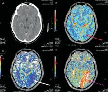
          Although noncontrast CT (NCCT) remains the most commonly used imaging modality to differentiate between ischemic and hemorrhagic stroke, to identify early CT changes, and to rule out stroke mimics, it is not sensitive enough to identify the infarct core or the mechanism of ischemic stroke. MRI, including magnetic resonance angiography (MRA), is the most useful imaging modality for the evaluation of acute stroke; it provides information about the mechanism as well as the vascular territory of the stroke. MRI also provides complete information about the status of tissue through diffusion-weighted imaging (DWI) and about arterial patency by means of MRA. DWI shows acute lesions within minutes of onset of ischemia, while MRA can evaluate extracranial as well as intracranial vessels Evaluation of the proportion of penumbra vs infarcted tissue is another issue to be considered when instituting thrombolysis, and its clinical usefulness is being assessed in a number of ongoing trials. Penumbral tissue can be identified by perfusion MRI. CT perfusion (CTP) is an emerging alternative, providing similar information about the penumbra and infarct core. A combined approach of NCCT, CT angiography (CTA), and CTP is now being employed at many centers and is known as multimodal CT imaging (MMCT). MMCT provides information about the pathophysiology of acute stroke which is comparable to that provided by MRI, and the technique has the potential to refine patient selection for thrombolysis.

          Related collections

          Most cited references107

          • Record: found
          • Abstract: found
          • Article: not found

          Global mortality, disability, and the contribution of risk factors: Global Burden of Disease Study.

          Prevention and control of disease and injury require information about the leading medical causes of illness and exposures or risk factors. The assessment of the public-health importance of these has been hampered by the lack of common methods to investigate the overall, worldwide burden. The Global Burden of Disease Study (GBD) provides a standardised approach to epidemiological assessment and uses a standard unit, the disability-adjusted life year (DALY), to aid comparisons. DALYs for each age-sex group in each GBD region for 107 disorders were calculated, based on the estimates of mortality by cause, incidence, average age of onset, duration, and disability severity. Estimates of the burden and prevalence of exposure in different regions of disorders attributable to malnutrition, poor water supply, sanitation and personal and domestic hygiene, unsafe sex, tobacco use, alcohol, occupation, hypertension, physical inactivity, use of illicit drugs, and air pollution were developed. Developed regions account for 11.6% of the worldwide burden from all causes of death and disability, and account for 90.2% of health expenditure worldwide. Communicable, maternal, perinatal, and nutritional disorders explain 43.9%; non-communicable causes 40.9%; injuries 15.1%; malignant neoplasms 5.1%; neuropsychiatric conditions 10.5%; and cardiovascular conditions 9.7% of DALYs worldwide. The ten leading specific causes of global DALYs are, in descending order, lower respiratory infections, diarrhoeal diseases, perinatal disorders, unipolar major depression, ischaemic heart disease, cerebrovascular disease, tuberculosis, measles, road-traffic accidents, and congenital anomalies. 15.9% of DALYs worldwide are attributable to childhood malnutrition and 6.8% to poor water, and sanitation and personal and domestic hygiene. The three leading contributors to the burden of disease are communicable and perinatal disorders affecting children. The substantial burdens of neuropsychiatric disorders and injuries are under-recognised. The epidemiological transition in terms of DALYs has progressed substantially in China, Latin America and the Caribbean, other Asia and islands, and the middle eastern crescent. If the burdens of disability and death are taken into account, our list differs substantially from other lists of the leading causes of death. DALYs provide a common metric to aid meaningful comparison of the burden of risk factors, diseases, and injuries.
            Bookmark
            • Record: found
            • Abstract: found
            • Article: not found

            Tissue Plasminogen Activator for Acute Ischemic Stroke

            (1996)
            Thrombolytic therapy for acute ischemic stroke has been approached cautiously because there were high rates of intracerebral hemorrhage in early clinical trials. We performed a randomized, double-blind trial of intravenous recombinant tissue plasminogen activator (t-PA) for ischemic stroke after recent pilot studies suggested that t-PA was beneficial when treatment was begun within three hours of the onset of stroke. The trial had two parts. Part 1 (in which 291 patients were enrolled) tested whether t-PA had clinical activity, as indicated by an improvement of 4 points over base-line values in the score of the National Institutes of Health stroke scale (NIHSS) or the resolution of the neurologic deficit within 24 hours of the onset of stroke. Part 2 (in which 333 patients were enrolled) used a global test statistic to assess clinical outcome at three months, according to scores on the Barthel index, modified Rankin scale, Glasgow outcome scale, and NIHSS: In part 1, there was no significant difference between the group given t-PA and that given placebo in the percentages of patients with neurologic improvement at 24 hours, although a benefit was observed for the t-PA group at three months for all four outcome measures. In part 2, the long-term clinical benefit of t-PA predicted by the results of part 1 was confirmed (global odds ratio for a favorable outcome, 1.7; 95 percent confidence interval, 1.2 to 2.6). As compared with patients given placebo, patients treated with t-PA were at least 30 percent more likely to have minimal or no disability at three months on the assessment scales. Symptomatic intracerebral hemorrhage within 36 hours after the onset of stroke occurred in 6.4 percent of patients given t-PA but only 0.6 percent of patients given placebo (P < 0.001). Mortality at three months was 17 percent in the t-PA group and 21 percent in the placebo group (P = 0.30). Despite an increased incidence of symptomatic intracerebral hemorrhage, treatment with intravenous t-PA within three hours of the onset of ischemic stroke improved clinical outcome at three months.
              Bookmark
              • Record: found
              • Abstract: found
              • Article: not found

              Probabilistic diffusion tractography with multiple fibre orientations: What can we gain?

              We present a direct extension of probabilistic diffusion tractography to the case of multiple fibre orientations. Using automatic relevance determination, we are able to perform online selection of the number of fibre orientations supported by the data at each voxel, simplifying the problem of tracking in a multi-orientation field. We then apply the identical probabilistic algorithm to tractography in the multi- and single-fibre cases in a number of example systems which have previously been tracked successfully or unsuccessfully with single-fibre tractography. We show that multi-fibre tractography offers significant advantages in sensitivity when tracking non-dominant fibre populations, but does not dramatically change tractography results for the dominant pathways.
                Bookmark

                Author and article information

                Journal
                Ann Indian Acad Neurol
                Ann Indian Acad Neurol
                AIAN
                Annals of Indian Academy of Neurology
                Medknow Publications & Media Pvt Ltd (India )
                0972-2327
                1998-3549
                January 2008
                : 11
                : Suppl 1
                : S12-S23
                Affiliations
                [1]National Stroke Research Institute, Austin Hospital, University of Melbourne, Heidelberg, 3081, Australia
                Author notes
                For correspondence: G. A. Donnan, National Stroke Research Institute, Austin Hospital, University of Melbourne, 300 Waterdale Road, Heidelberg Heights, Victoria3081, Australia. E-mail: gdonnan@ 123456unimelb.edu.au
                Article
                AIAN-11-12
                10.4103/0972-2327.41715
                9204117
                ffabeed8-1e7f-416e-bbfa-e77c53f8c7ba
                Copyright: © 2008 Annals of Indian Academy of Neurology

                This is an open access journal, and articles are distributed under the terms of the Creative Commons Attribution-NonCommercial-ShareAlike 4.0 License, which allows others to remix, tweak, and build upon the work non-commercially, as long as appropriate credit is given and the new creations are licensed under the identical terms.

                History
                : 03 December 2007
                : 28 March 2008
                : 29 April 2008
                Categories
                Review Article

                Neurology
                imaging,stroke,thrombolysis,penumbra,infarct core,multi model ct stroke imaging,perfusion/diffusion mri

                Comments

                Comment on this article