0
views
0
recommends
+1 Recommend
0 collections
    0
    shares
      • Record: found
      • Abstract: found
      • Article: found
      Is Open Access

      The added value of conventional defecography and MRI defecography in clinical decision making on treatment for posterior compartment prolapse

      research-article

      Read this article at

      Bookmark
          There is no author summary for this article yet. Authors can add summaries to their articles on ScienceOpen to make them more accessible to a non-specialist audience.

          Abstract

          Introduction and hypothesis

          Conventional defecography and MRI defecography can be requested as an additional test for diagnosing and differentiating the type of posterior compartment prolapse and/or obstructive defecation disorders. The objective of this study was to determine the added value of conventional defecography, conventional defecography and MRI defecography for clinical decision-making on treatment for patients with posterior compartment prolapse.

          Methods

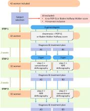
          Four gynecologists were asked to fill in their treatment plan per patient for 32 cases for three different steps. Step 1 consisted of information on the anamnesis and physical examination (POP-Q). Step 2 consisted of Step 1, including conventional defecography (group A) or MRI defecography (group B). In Step 3, all gynecologists received the information on Step 1 including both conventional defecography and MRI defecography. Data analysis solely focused on the assessment of changes in the gynecological treatment plan of the posterior compartment.

          Results

          After Step 2 a change in treatment plan occurred in 37% and 48% of the women in groups A and B, respectively. Accordingly, after Step 3 (including all imaging data), a change in treatment plan occurred in 19% and 52% of the women in groups A and B, respectively. A change within the surgery group (when a different type of surgery was selected) was seen for a total of 11 cases in group A and 20 in group B in all steps combined.

          Conclusions

          Both conventional defecography and MRI defecography had an large effect on the treatment plan for patients with posterior compartment prolapse. The dedicated added value of the imaging modality individually cannot be concluded yet.

          Related collections

          Most cited references11

          • Record: found
          • Abstract: found
          • Article: not found

          MR imaging-based assessment of the female pelvic floor.

          Pelvic floor weakness is a functional condition that affects the anatomic structures supporting the pelvic organs: fasciae, ligaments, and muscles. It is a prevalent disorder among people older than 50 years, especially women, and may substantially diminish their quality of life. Many complex causes of pelvic floor weakness have been described, but the greatest risk factors are aging and female sex. Pelvic floor weakness can provoke a wide range of symptoms, including pain, urinary and fecal incontinence, constipation, difficulty in voiding, a sense of pressure, and sexual dysfunction. When the condition is diagnosed solely on the basis of physical and clinical examination, the compartments involved and the site of prolapse are frequently misidentified. Such errors contribute to a high number of failed interventions. Magnetic resonance (MR) imaging, which allows visualization of all three compartments, has proved a reliable technique for accurate diagnosis, especially when involvement of multiple compartments is suspected. MR imaging allows precise evaluation of ligaments, muscles, and pelvic organs and provides accurate information for appropriate surgical treatment. Moreover, dynamic MR imaging with steady-state sequences enables the evaluation of functional disorders of the pelvic floor. The authors review the pelvic floor anatomy, describe the MR imaging protocol used in their institutions, survey common MR imaging findings in the presence of pelvic floor weakness, and highlight key details that radiologists should provide surgeons to ensure effective treatment and improved outcomes.
            Bookmark
            • Record: found
            • Abstract: found
            • Article: not found

            Nonsurgical management of pelvic organ prolapse.

            Although surgical management of symptomatic pelvic organ prolapse (POP) is common and often necessary, conservative treatments such as pessaries, pelvic floor muscle training, or both can usually result in symptomatic improvement. When treating patients with POP, health care practitioners should focus primarily on identification and alleviation of POP-related symptoms. It is appropriate to offer nonsurgical management to most people with POP. This article reviews the objective and subjective evaluation and nonsurgical management of POP, emphasizing a simple, practical approach to pessary fitting and management.
              Bookmark
              • Record: found
              • Abstract: found
              • Article: not found

              Dynamic MR defecography of the posterior compartment: Indications, techniques and MRI features.

              Pelvic floor weakness is characterized by abnormal symptomatic displacement of pelvic organs. It represents a complex clinical problem most commonly seen in middle-aged and elderly parous women. Its diagnosis remains difficult in many cases, since these disorders typically present with nonspecific symptoms, such as pelvic pain, incontinence and constipation. Fluoroscopic colpocystodefecography has been proven to surpass physical examination in the detection and characterization of functional abnormalities of the anorectum and surrounding pelvic structures. Similarly, MR defecography, performed either with an open- or closed-configuration unit, appears to be an accurate imaging technique to assess clinically relevant pelvic floor abnormalities. Moreover, MR defecography negates the need to expose the patient to harmful ionizing radiation and allows excellent depiction of the surrounding soft tissues of the pelvis. In this manuscript, we review the techniques and indications of MR defecography, and illustrate the MRI features of a vast array of morphologic and functional pelvic floor disorders, with emphasis on the posterior pelvic compartment (anorectum).
                Bookmark

                Author and article information

                Contributors
                a.t.m.grob@utwente.nl
                Journal
                Int Urogynecol J
                Int Urogynecol J
                International Urogynecology Journal
                Springer International Publishing (Cham )
                0937-3462
                1433-3023
                11 April 2022
                11 April 2022
                2023
                : 34
                : 2
                : 507-515
                Affiliations
                [1 ]GRID grid.6214.1, ISNI 0000 0004 0399 8953, Multi-Modality Medical Imaging, Faculty of Science and Technology, Technical Medical Centre, , University of Twente, ; Building Technohal, Drienerlolaan 5, 7522 NB Enschede, The Netherlands
                [2 ]GRID grid.417370.6, ISNI 0000 0004 0502 0983, Department of Obstetrics and Gynecology, , Ziekenhuisgroep Twente, ; Hengelo, The Netherlands
                [3 ]GRID grid.417370.6, ISNI 0000 0004 0502 0983, Department of Radiology, , Ziekenhuisgroep Twente, ; Hengelo, The Netherlands
                Article
                5181
                10.1007/s00192-022-05181-x
                9870817
                35403883
                feedb04a-aff7-4b6e-9475-8780ed7d4e0e
                © The Author(s) 2022

                Open AccessThis article is licensed under a Creative Commons Attribution 4.0 International License, which permits use, sharing, adaptation, distribution and reproduction in any medium or format, as long as you give appropriate credit to the original author(s) and the source, provide a link to the Creative Commons licence, and indicate if changes were made. The images or other third party material in this article are included in the article’s Creative Commons licence, unless indicated otherwise in a credit line to the material. If material is not included in the article’s Creative Commons licence and your intended use is not permitted by statutory regulation or exceeds the permitted use, you will need to obtain permission directly from the copyright holder. To view a copy of this licence, visit http://creativecommons.org/licenses/by/4.0/.

                History
                : 29 November 2021
                : 4 March 2022
                Categories
                Original Article
                Custom metadata
                © The International Urogynecological Association 2023

                Obstetrics & Gynecology
                clinical decision-making,mri defecography,prolapse,conventional defecography

                Comments

                Comment on this article