35
views
0
recommends
+1 Recommend
0 collections
    0
    shares
      • Record: found
      • Abstract: found
      • Article: found
      Is Open Access

      Lipidomic study of kidney in a mouse model with urine flow obstruction

      research-article

      Read this article at

      Bookmark
          There is no author summary for this article yet. Authors can add summaries to their articles on ScienceOpen to make them more accessible to a non-specialist audience.

          Abstract

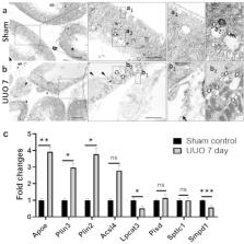
          Obstructed urine flow is known to cause structural and functional kidney damage leading to renal fibrosis. However, limited information is available on the change in kidney lipids during urinary tract obstruction. In this study, we investigated the change in lipidome in a mouse model with unilateral ureteral obstruction (UUO). The establishment of the UUO model was confirmed by histopathological examination using transmission electron microscopy. Untargeted liquid chromatography/mass spectrometry was carried out over a time course of 4 and 7 days. Compared to the sham control, the UUO kidney at 7 days showed dilatation of the renal tubule with loss of brush borders and thickening of the capillary endothelium. In the kidney lipidomes obtained from the UUO 7 days group compared to the control, a significant decrease of ceramide, sphingomyelin, phosphatidylcholine, lysophospholipids, and phosphatidylethanolamine was observed, whereas cholesteryl esters, free fatty acids, phosphatidylglycerol, and cardiolipins were significantly increased. The present study revealed the disturbed lipid metabolism in the UUO model, which may provide a clue to potential lipid pathways and therapeutic targets for the early stage of renal fibrosis.

          Related collections

          Most cited references60

          • Record: found
          • Abstract: found
          • Article: not found

          The distribution and function of phosphatidylserine in cellular membranes.

          Phosphatidylserine (PS) is the most abundant negatively charged phospholipid in eukaryotic membranes. PS directs the binding of proteins that bear C2 or gamma-carboxyglutamic domains and contributes to the electrostatic association of polycationic ligands with cellular membranes. Rather than being evenly distributed, PS is found preferentially in the inner leaflet of the plasma membrane and in endocytic membranes. The loss of PS asymmetry is an early indicator of apoptosis and serves as a signal to initiate blood clotting. This review discusses the determinants and functional implications of the subcellular distribution and membrane topology of PS.
            Bookmark
            • Record: found
            • Abstract: found
            • Article: not found

            Exposure of phosphatidylserine on the cell surface.

            Phosphatidylserine (PtdSer) is a phospholipid that is abundant in eukaryotic plasma membranes. An ATP-dependent enzyme called flippase normally keeps PtdSer inside the cell, but PtdSer is exposed by the action of scramblase on the cell's surface in biological processes such as apoptosis and platelet activation. Once exposed to the cell surface, PtdSer acts as an 'eat me' signal on dead cells, and creates a scaffold for blood-clotting factors on activated platelets. The molecular identities of the flippase and scramblase that work at plasma membranes have long eluded researchers. Indeed, their identity as well as the mechanism of the PtdSer exposure to the cell surface has only recently been revealed. Here, we describe how PtdSer is exposed in apoptotic cells and in activated platelets, and discuss PtdSer exposure in other biological processes.
              Bookmark
              • Record: found
              • Abstract: found
              • Article: not found

              Phosphatidylethanolamine Metabolism in Health and Disease.

              Phosphatidylethanolamine (PE) is the second most abundant glycerophospholipid in eukaryotic cells. The existence of four only partially redundant biochemical pathways that produce PE, highlights the importance of this essential phospholipid. The CDP-ethanolamine and phosphatidylserine decarboxylase pathways occur in different subcellular compartments and are the main sources of PE in cells. Mammalian development fails upon ablation of either pathway. Once made, PE has diverse cellular functions that include serving as a precursor for phosphatidylcholine and a substrate for important posttranslational modifications, influencing membrane topology, and promoting cell and organelle membrane fusion, oxidative phosphorylation, mitochondrial biogenesis, and autophagy. The importance of PE metabolism in mammalian health has recently emerged following its association with Alzheimer's disease, Parkinson's disease, nonalcoholic liver disease, and the virulence of certain pathogenic organisms.
                Bookmark

                Author and article information

                Contributors
                masum@sau.edu.bd
                keino@hs.hokudai.ac.jp
                Journal
                Sci Rep
                Sci Rep
                Scientific Reports
                Nature Publishing Group UK (London )
                2045-2322
                5 August 2024
                5 August 2024
                2024
                : 14
                : 18042
                Affiliations
                [1 ]Faculty of Health Sciences, Hokkaido University, ( https://ror.org/02e16g702) Kita-12 Nishi-5, Kita-ku, Sapporo, 060-0812 Japan
                [2 ]Department of Anatomy, Histology and Physiology, Faculty of Animal Science and Veterinary Medicine, Sher-e-Bangla Agricultural University, ( https://ror.org/03ht0cf17) Dhaka, 1207 Bangladesh
                [3 ]Laboratory of Anatomy, Department of Basic Veterinary Medicine, Faculty of Veterinary Medicine, Hokkaido University, ( https://ror.org/02e16g702) Sapporo, 060-0818 Japan
                [4 ]Graduate School of Global Food Resources, Hokkaido University, ( https://ror.org/02e16g702) Kita 9, Nishi 9, Kita-ku, Sapporo, 060-0809 Japan
                [5 ]Departments of Physiology, Medicine, Molecular Biology Immunology and Biochemistry, and Pharmaceutical Sciences, University of Tennessee Health Science Center, ( https://ror.org/0011qv509) Memphis, TN 38163 USA
                [6 ]Laboratory of Agrobiomedical Science, Faculty of Agriculture, Hokkaido University, ( https://ror.org/02e16g702) Sapporo, 060-0809 Japan
                [7 ]Department of Nutrition, Sapporo University of Health Sciences, ( https://ror.org/04bz6gw73) Nakanuma, Nishi-4-3-1-15, Higashi-ku, Sapporo, 007-0894 Japan
                Article
                68270
                10.1038/s41598-024-68270-5
                11298537
                39098953
                fd265f2f-e418-4ea0-bdd2-9159fee74ef6
                © The Author(s) 2024

                Open Access This article is licensed under a Creative Commons Attribution-NonCommercial-NoDerivatives 4.0 International License, which permits any non-commercial use, sharing, distribution and reproduction in any medium or format, as long as you give appropriate credit to the original author(s) and the source, provide a link to the Creative Commons licence, and indicate if you modified the licensed material. You do not have permission under this licence to share adapted material derived from this article or parts of it. The images or other third party material in this article are included in the article’s Creative Commons licence, unless indicated otherwise in a credit line to the material. If material is not included in the article’s Creative Commons licence and your intended use is not permitted by statutory regulation or exceeds the permitted use, you will need to obtain permission directly from the copyright holder. To view a copy of this licence, visit http://creativecommons.org/licenses/by-nc-nd/4.0/.

                History
                : 9 August 2023
                : 22 July 2024
                Funding
                Funded by: Japanese Society for the Promotion of Science KAKENHI
                Award ID: 19H0311719
                Award ID: 19K0786109
                Award ID: 22K1485002
                Award ID: 21K1481201
                Award ID: 19F19092
                Award Recipient :
                Categories
                Article
                Custom metadata
                © Springer Nature Limited 2024

                Uncategorized
                lipid,liquid chromatography,mass spectrometry,kidney injury,unilateral ureteral obstruction,renal tubulointerstitial lesion,kidney,diagnostic markers,bioanalytical chemistry

                Comments

                Comment on this article