23
views
0
recommends
+1 Recommend
0 collections
    0
    shares
      • Record: found
      • Abstract: found
      • Article: found
      Is Open Access

      Prognostic evaluation of esophageal cancer patients with stages I-III

      research-article

      Read this article at

      Bookmark
          There is no author summary for this article yet. Authors can add summaries to their articles on ScienceOpen to make them more accessible to a non-specialist audience.

          Abstract

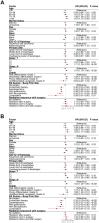
          Purpose: The purpose of this study was to investigate the impact of clinicopathological factors and treatments on the overall survival (OS) and esophageal cancer-specific survival (ECSS) of stages I-III esophageal cancer (EC) patients and to establish a prognostic visual nomogram. Methods: We collected clinical data of patients diagnosed with stages I-III EC without receiving chemotherapy from 2004 to 2014 from the Surveillance, Epidemiology, and End Results (SEER) database. Prognoses were analyzed using the R language software, and nomograms were obtained according to the visual processing logistic regression model, which was verified using the Harrell C-index, receiver operating characteristic (ROC) curve, and calibration curve. Results: A total of 4,305 patients were selected, mostly white males. Most patients were over 60 years old and old age predicted poor prognosis. EC, primarily adenocarcinoma, occurred mostly in the lower third of the esophagus. About half of the patients had T1 (58.00%) and grade II (50.41%) cancer. Of all the patients, 2,448 was treated with surgery and the majority (n = 1,476; 64.85%) of these patients had stage I EC. Stages I-III patients underwent surgery had significantly better OS and ECSS, and endoscopic therapy was associated with the best outcome amongst all the surgical methods. 3.67% of the patients received radiotherapy, predominantly postoperative radiotherapy (2.69%). Older age, squamous cell carcinoma, overlapping lesion of the esophagus, and grades II and III were high-risk factors for poor OS and ECSS for stage I patients, whereas endoscopic therapy, esophagectomy, and esophagectomy with gastrectomy were low-risk factors. Stage II patients with older age, male sex, T3, N1, and grades II and III had shorter OS and ECSS, but patients with any surgical treatment had significantly longer OS and ECSS. T4, N1, and grade III correlated negatively with OS and ECSS in stage III patients, and any surgical treatment correlated positively with longer OS and ECSS. The OS and ECSS rates of stages I-III EC patients with a total score of more than 150 points in the nomogram were both only 40% after 3 years and 30% after 5 years. The C-index, ROC curve, and calibration curve indicated that the nomograms established in this study were suitable to assess patient prognosis. Conclusion: The nomogram established in this study is an effective clinical tool to predict the prognosis of stages I-III EC patients without chemotherapy.

          Related collections

          Most cited references23

          • Record: found
          • Abstract: found
          • Article: not found

          Marital status and survival in patients with cancer.

          To examine the impact of marital status on stage at diagnosis, use of definitive therapy, and cancer-specific mortality among each of the 10 leading causes of cancer-related death in the United States.
            Bookmark
            • Record: found
            • Abstract: found
            • Article: not found

            Development and validation of a prognostic nomogram for recurrence-free survival after complete surgical resection of localised primary gastrointestinal stromal tumour: a retrospective analysis.

            Adjuvant imatinib mesylate prolongs recurrence-free survival (RFS) after resection of localised primary gastrointestinal stromal tumours (GIST). We aimed to develop a nomogram to predict RFS after surgery in the absence of adjuvant therapy to help guide patient selection for adjuvant imatinib therapy. A nomogram to predict RFS based on tumour size (cm), location (stomach, small intestine, colon/rectum, or other), and mitotic index ( or =5 mitoses per 50 high-power fields) was developed from 127 patients treated at Memorial Sloan-Kettering Cancer Center (MSKCC), New York, NY, USA. The nomogram was tested in patients from the Spanish Group for Research on Sarcomas (GEIS; n=212) and the Mayo Clinic, Rochester, MN, USA (Mayo; n=148). The nomogram was assessed by calculating concordance probabilities and testing calibration of predicted RFS with observed RFS. Concordance probabilities were also compared with those of three commonly used staging systems. The nomogram had a concordance probability of 0.78 (SE 0.02) in the MSKCC dataset, and 0.76 (0.03) and 0.80 (0.02) in the validation cohorts. Nomogram predictions were well calibrated. Inclusion of tyrosine kinase mutation status in the nomogram did not improve its discriminatory ability. Concordance probabilities of the nomogram were better than those of the two NIH staging systems (0.76 [0.03] vs 0.70 [0.04, p=0.04] and 0.66 [0.04, p=0.01] in the GEIS validation cohort; 0.80 [0.02] vs 0.74 [0.02, p=0.04] and 0.78 [0.02, p=0.05] in the Mayo cohort) and similar to those of the AFIP-Miettinen staging system (0.76 [0.03] vs 0.73 [0.004, p=0.28] in the GEIS cohort; 0.80 [0.02] vs 0.76 [0.003, p=0.09] in the Mayo cohort). Nomogram predictions of RFS seemed better calibrated than predictions made with the AFIP-Miettinen system. The nomogram accurately predicts RFS after resection of localised primary GIST and could be used to select patients for adjuvant imatinib therapy.
              Bookmark
              • Record: found
              • Abstract: found
              • Article: not found

              Long-term results and risk factor analysis for recurrence after curative endoscopic therapy in 349 patients with high-grade intraepithelial neoplasia and mucosal adenocarcinoma in Barrett's oesophagus.

              Endoscopic therapy is increasingly being used in the treatment of high-grade intraepithelial neoplasia (HGIN) and mucosal adenocarcinoma (BC) in patients with Barrett's oesophagus. This report provides 5 year follow-up data from a large prospective study investigating the efficacy and safety of endoscopic treatment in these patients and analysing risk factors for recurrence. Prospective case series. Academic tertiary care centre. Between October 1996 and September 2002, 61 patients with HGIN and 288 with BC were included (173 with short-segment and 176 with long-segment Barrett's oesophagus) from a total of 486 patients presenting with Barrett's neoplasia. Patients with submucosal or more advanced cancer were excluded. Endoscopic therapy. Rate of complete remission and recurrence rate, tumour-associated death. Endoscopic resection was performed in 279 patients, photodynamic therapy in 55, and both procedures in 13; two patients received argon plasma coagulation. The mean follow-up period was 63.6 (SD 23.1) months. Complete response (CR) was achieved in 337 patients (96.6%); surgery was necessary in 13 (3.7%) after endoscopic therapy failed. Metachronous lesions developed during the follow-up in 74 patients (21.5%); 56 died of concomitant disease, but none died of BC. The calculated 5 year survival rate was 84%. The risk factors most frequently associated with recurrence were piecemeal resection, long-segment Barrett's oesophagus, no ablative therapy of Barrett's oesophagus after CR, time until CR achieved >10 months and multifocal neoplasia. This study showed that endoscopic therapy was highly effective and safe, with an excellent long-term survival rate. The risk factors identified may help stratify patients who are at risk for recurrence and those requiring more intensified follow-up.
                Bookmark

                Author and article information

                Journal
                Aging (Albany NY)
                Aging (Albany NY)
                Aging
                Aging (Albany NY)
                Impact Journals
                1945-4589
                31 July 2020
                23 July 2020
                : 12
                : 14
                : 14736-14753
                Affiliations
                [1 ]Division of Gastroenterology, Liyuan Hospital, Tongji Medical College, Huazhong University of Science and Technology, Wuhan 430077, China
                [2 ]Cancer Center, Union Hospital, Tongji Medical College, Huazhong University of Science and Technology, Wuhan 430022, China
                [3 ]Yangchunhu Community Hospital, Liyuan Hospital, Tongji Medical College, Huazhong University of Science and Technology, Wuhan 430077, China
                Author notes
                Correspondence to: Zao-zao Huang; email: huangzaozao1986@163.com
                Correspondence to: Zhi-fan Xiong; email: 1992ly0503@hust.edu.cn
                Article
                103532 103532
                10.18632/aging.103532
                7425498
                32701062
                fcdb3bef-c89f-4da8-90ee-4acc155aee2a
                Copyright © 2020 Qiu et al.

                This is an open-access article distributed under the terms of the Creative Commons Attribution License (CC BY 3.0), which permits unrestricted use, distribution, and reproduction in any medium, provided the original author and source are credited.

                History
                : 17 March 2020
                : 09 June 2020
                Categories
                Research Paper

                Cell biology
                esophageal cancer,seer database,surgery,nomogram,prognosis
                Cell biology
                esophageal cancer, seer database, surgery, nomogram, prognosis

                Comments

                Comment on this article