17
views
0
recommends
+1 Recommend
0 collections
    0
    shares
      • Record: found
      • Abstract: found
      • Article: found
      Is Open Access

      “Switchboard” malfunction in motor neuron diseases: Selective pathology of thalamic nuclei in amyotrophic lateral sclerosis and primary lateral sclerosis

      research-article

      Read this article at

      Bookmark
          There is no author summary for this article yet. Authors can add summaries to their articles on ScienceOpen to make them more accessible to a non-specialist audience.

          Abstract

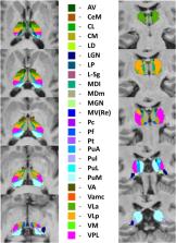
          The thalamus is a key cerebral hub relaying a multitude of corticoefferent and corticoafferent connections and mediating distinct extrapyramidal, sensory, cognitive and behavioural functions. While the thalamus consists of dozens of anatomically well-defined nuclei with distinctive physiological roles, existing imaging studies in motor neuron diseases typically evaluate the thalamus as a single structure. Based on the unique cortical signatures observed in ALS and PLS, we hypothesised that similarly focal thalamic involvement may be observed if the nuclei are individually evaluated. A prospective imaging study was undertaken with 100 patients with ALS, 33 patients with PLS and 117 healthy controls to characterise the integrity of thalamic nuclei. ALS patients were further stratified for the presence of GGGGCC hexanucleotide repeat expansions in C9orf72. The thalamus was segmented into individual nuclei to examine their volumetric profile. Additionally, thalamic shape deformations were evaluated by vertex analyses and focal density alterations were examined by region-of-interest morphometry. Our data indicate that C9orf72 negative ALS patients and PLS patients exhibit ventral lateral and ventral anterior involvement, consistent with the ‘motor’ thalamus. Degeneration of the sensory nuclei was also detected in C9orf72 negative ALS and PLS. Both ALS groups and the PLS cohort showed focal changes in the mediodorsal-paratenial-reuniens nuclei, which mediate memory and executive functions. PLS patients exhibited distinctive thalamic changes with marked pulvinar and lateral geniculate atrophy compared to both controls and C9orf72 negative ALS. The considerable ventral lateral and ventral anterior pathology detected in both ALS and PLS support the emerging literature of extrapyramidal dysfunction in MND. The involvement of sensory nuclei is consistent with sporadic reports of sensory impairment in MND. The unique thalamic signature of PLS is in line with the distinctive clinical features of the phenotype. Our data confirm phenotype-specific patterns of thalamus involvement in motor neuron diseases with the preferential involvement of nuclei mediating motor and cognitive functions. Given the selective involvement of thalamic nuclei in ALS and PLS, future biomarker and natural history studies in MND should evaluate individual thalamic regions instead overall thalamic changes.

          Related collections

          Most cited references85

          • Record: found
          • Abstract: not found
          • Article: not found

          Thalamus plays a central role in ongoing cortical functioning

            Bookmark
            • Record: found
            • Abstract: found
            • Article: found
            Is Open Access

            A probabilistic atlas of the human thalamic nuclei combining ex vivo MRI and histology

            The human thalamus is a brain structure that comprises numerous, highly specific nuclei. Since these nuclei are known to have different functions and to be connected to different areas of the cerebral cortex, it is of great interest for the neuroimaging community to study their volume, shape and connectivity in vivo with MRI. In this study, we present a probabilistic atlas of the thalamic nuclei built using ex vivo brain MRI scans and histological data, as well as the application of the atlas to in vivo MRI segmentation. The atlas was built using manual delineation of 26 thalamic nuclei on the serial histology of 12 whole thalami from six autopsy samples, combined with manual segmentations of the whole thalamus and surrounding structures (caudate, putamen, hippocampus, etc.) made on in vivo brain MR data from 39 subjects. The 3D structure of the histological data and corresponding manual segmentations was recovered using the ex vivo MRI as reference frame, and stacks of blockface photographs acquired during the sectioning as intermediate target. The atlas, which was encoded as an adaptive tetrahedral mesh, shows a good agreement with previous histological studies of the thalamus in terms of volumes of representative nuclei. When applied to segmentation of in vivo scans using Bayesian inference, the atlas shows excellent test-retest reliability, robustness to changes in input MRI contrast, and ability to detect differential thalamic effects in subjects with Alzheimer's disease. The probabilistic atlas and companion segmentation tool are publicly available as part of the neuroimaging package FreeSurfer.
              Bookmark
              • Record: found
              • Abstract: found
              • Article: not found

              Functional-anatomical validation and individual variation of diffusion tractography-based segmentation of the human thalamus.

              Parcellation of the human thalamus based on cortical connectivity information inferred from non-invasive diffusion-weighted images identifies sub-regions that we have proposed correspond to nuclei. Here we test the functional and anatomical validity of this proposal by comparing data from diffusion tractography, cytoarchitecture and functional imaging. We acquired diffusion imaging data in eleven healthy subjects and performed probabilistic tractography from voxels within the thalamus. Cortical connectivity information was used to divide the thalamus into sub-regions with highest probability of connectivity to distinct cortical areas. The relative volumes of these connectivity-defined sub-regions correlate well with volumetric predictions based on a histological atlas. Previously reported centres of functional activation within the thalamus during motor or executive tasks co-localize within atlas regions showing high probabilities of connection to motor or prefrontal cortices, respectively. This work provides a powerful validation of quantitative grey matter segmentation using diffusion tractography in humans. Co-registering thalamic sub-regions from 11 healthy individuals characterizes inter-individual variation in segmentation and results in a population-based atlas of the human thalamus that can be used to assign likely anatomical labels to thalamic locations in standard brain space. This provides a tool for specific localization of functional activations or lesions to putative thalamic nuclei.
                Bookmark

                Author and article information

                Contributors
                Journal
                Neuroimage Clin
                Neuroimage Clin
                NeuroImage : Clinical
                Elsevier
                2213-1582
                30 May 2020
                2020
                30 May 2020
                : 27
                : 102300
                Affiliations
                [a ]Computational Neuroimaging Group, Biomedical Sciences Institute, Trinity College Dublin, Ireland
                [b ]Department of Neurology, Aeginition Hospital, University of Athens, Greece
                [c ]Electronics and Computer Science, University of Southampton, United Kingdom
                [d ]Complex Trait Genomics Laboratory, Smurfit Institute of Genetics, Trinity College Dublin, Ireland
                [e ]Department of Psychology, Beaumont Hospital Dublin, Ireland
                [f ]Department of Neurology, St James’s Hospital, Dublin, Ireland
                [g ]Department of Neurology, Western Health & Social Care Trust, Belfast, United Kingdom
                Author notes
                [* ]Corresponding author at: Room 5.43, Computational Neuroimaging Group, Trinity Biomedical Sciences Institute, Trinity College Dublin, Pearse Street, Dublin 2, Ireland. bedep@ 123456tcd.ie
                Article
                S2213-1582(20)30137-6 102300
                10.1016/j.nicl.2020.102300
                7303672
                32554322
                fc83a91a-3b47-4e48-9d80-2bcf01566fa8
                © 2020 The Authors

                This is an open access article under the CC BY-NC-ND license (http://creativecommons.org/licenses/by-nc-nd/4.0/).

                History
                : 12 February 2020
                : 22 May 2020
                : 23 May 2020
                Categories
                Regular Article

                motor neuron disease,amyotrophic lateral sclerosis,primary lateral sclerosis,thalamus,thalamic nuclei,neuroimaging

                Comments

                Comment on this article