63
views
0
recommends
+1 Recommend
0 collections
    0
    shares
      • Record: found
      • Abstract: found
      • Article: found
      Is Open Access

      Cellular Expression Profile for Interstitial Cells of Cajal in Bladder - A Cell Often Misidentified as Myocyte or Myofibroblast

      research-article
      1 , 2 , * , 1 , 2 , 1 , 3
      PLoS ONE
      Public Library of Science

      Read this article at

      Bookmark
          There is no author summary for this article yet. Authors can add summaries to their articles on ScienceOpen to make them more accessible to a non-specialist audience.

          Abstract

          Background

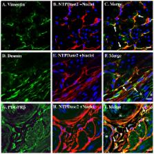
          Interstitial cells of Cajal (ICC) have been identified in urinary bladder of several species, but their presence in mice remains uncertain. Meanwhile, dozens of reports indicate that dysregulation of connexin 43 plays an important role in bladder overactivity, but its localization has not been clearly defined, with reports of expression in either the smooth muscle or in myofibroblasts. We recently identified a population of ectonucleoside triphosphate diphosphohydrolase 2 (NTPDase2) positive cells that resemble ICC and are distinct from smooth muscle, fibroblasts, myofibroblasts and neurons. Thus we sought to define more clearly the molecular signature of ICC and in doing so resolve some of these uncertainties.

          Principle findings

          Immunofluorescent localization revealed that NTPDase2-positive cells lie closely adjacent to smooth muscle but are separate from them. NTPDase2 positive cells exhibited co-localization with the widely accepted ICC marker - c-kit. They were further shown to co-localize with other ICC markers CD34 and Ano1, but not with mast cell marker tryptase. Significantly, they show convincing co-localization with connexin 43, which was not present in smooth muscle. The identity of these cells as ICC was further confirmed by the presence of three mesenchymal markers – vimentin, desmin, and PDGFβ receptor, which indicates their mesenchymal origin. Finally, we observed for the first time, the presence of merlin/neurofibromin 2 in ICC. Normally considered a neuronal protein, the presence of merlin suggests ICC in bladder may have a role in neurotransmission.

          Conclusions

          NTPDase2 positive cells in mice bladder are ICC, which can be defined by the presence of c-Kit, CD34, Ano1, NTPDase2, connexin 43, vimentin, desmin, PDGFβ receptor and merlin/NF2. These data establish a definitive molecular expression profile, which can be used to assist in explorations of their functional roles, and the presence of NTPDase2 suggests that purinergic signaling plays a role in regulation of ICC function.

          Related collections

          Most cited references56

          • Record: found
          • Abstract: found
          • Article: not found

          Ano1 is a selective marker of interstitial cells of Cajal in the human and mouse gastrointestinal tract.

          Populations of interstitial cells of Cajal (ICC) are altered in several gastrointestinal neuromuscular disorders. ICC are identified typically by ultrastructure and expression of Kit (CD117), a protein that is also expressed on mast cells. No other molecular marker currently exists to independently identify ICC. The expression of ANO1 (DOG1, TMEM16A), a Ca(2+)-activated Cl(-) channel, in gastrointestinal stromal tumors suggests it may be useful as an ICC marker. The aims of this study were therefore to determine the distribution of Ano1 immunoreactivity compared with Kit and to establish whether Ano1 is a reliable marker for human and mouse ICC. Expression of Ano1 in human and mouse stomach, small intestine, and colon was investigated by immunofluorescence labeling using antibodies to Ano1 alone and in combination with antibodies to Kit. Colocalization of immunoreactivity was demonstrated by epifluorescence and confocal microscopy. In the muscularis propria, Ano1 immunoreactivity was restricted to cells with the morphology and distribution of ICC. All Ano1-positive cells in the muscularis propria were also Kit positive. Kit-expressing mast cells were not Ano1 positive. Some non-ICC in the mucosa and submucosa of human tissues were Ano1 positive but Kit negative. A few (3.2%) Ano1-positive cells in the human gastric muscularis propria were labeled weakly for Kit. Ano1 labels all classes of ICC and represents a highly specific marker for studying the distribution of ICC in mouse and human tissues with an advantage over Kit since it does not label mast cells.
            Bookmark
            • Record: found
            • Abstract: found
            • Article: not found

            Vesicular and conductive mechanisms of nucleotide release.

            Extracellular nucleotides and nucleosides promote a vast range of physiological responses, via activation of cell surface purinergic receptors. Virtually all tissues and cell types exhibit regulated release of ATP, which, in many cases, is accompanied by the release of uridine nucleotides. Given the relevance of extracellular nucleotide/nucleoside-evoked responses, understanding how ATP and other nucleotides are released from cells is an important physiological question. By facilitating the entry of cytosolic nucleotides into the secretory pathway, recently identified vesicular nucleotide and nucleotide-sugar transporters contribute to the exocytotic release of ATP and UDP-sugars not only from endocrine/exocrine tissues, but also from cell types in which secretory granules have not been biochemically characterized. In addition, plasma membrane connexin hemichannels, pannexin channels, and less-well molecularly defined ATP conducting anion channels have been shown to contribute to the release of ATP (and UTP) under a variety of conditions.
              Bookmark
              • Record: found
              • Abstract: found
              • Article: not found

              NF2 deficiency promotes tumorigenesis and metastasis by destabilizing adherens junctions.

              Mutation of the Neurofibromatosis 2 (NF2) tumor suppressor gene leads to cancer development in humans and mice. Recent studies suggest that Nf2 loss also contributes to tumor metastasis. The Nf2-encoded protein, merlin, is related to the ERM (ezrin, radixin, and moesin) family of membrane:cytoskeleton-associated proteins. However, the cellular mechanism whereby merlin controls cell proliferation from this location is not known. Here we show that the major cellular consequence of Nf2 deficiency in primary cells is an inability to undergo contact-dependent growth arrest and to form stable cadherin-containing cell:cell junctions. Merlin colocalizes and interacts with adherens junction (AJ) components in confluent wild-type cells, suggesting that the lack of AJs and contact-dependent growth arrest in Nf2(-/-) cells directly results from the absence of merlin at sites of cell:cell contact. Our studies indicate that merlin functions as a tumor and metastasis suppressor by controlling cadherin-mediated cell:cell contact.
                Bookmark

                Author and article information

                Contributors
                Role: Editor
                Journal
                PLoS One
                PLoS ONE
                plos
                plosone
                PLoS ONE
                Public Library of Science (San Francisco, USA )
                1932-6203
                2012
                7 November 2012
                : 7
                : 11
                : e48897
                Affiliations
                [1 ]Laboratory of Voiding Dysfunction, Department of Medicine, Beth Israel Deaconess Medical Center, and Harvard Medical School, Boston, Massachusetts, United States of America
                [2 ]Division of Nephrology, Department of Medicine, Beth Israel Deaconess Medical Center, and Harvard Medical School, Boston, Massachusetts, United States of America
                [3 ]Division of Matrix Biology; Department of Medicine, Beth Israel Deaconess Medical Center, and Harvard Medical School, Boston, Massachusetts, United States of America
                Centro Cardiologico Monzino, Italy
                Author notes

                Competing Interests: The authors have declared that no competing interests exist.

                Conceived and designed the experiments: WY MLZ WGH. Performed the experiments: WY. Analyzed the data: WY MLZ WGH. Contributed reagents/materials/analysis tools: WY MLZ WGH. Wrote the paper: WY MLZ WGH. NA.

                Article
                PONE-D-12-18088
                10.1371/journal.pone.0048897
                3492220
                23145014
                fae6c20f-3240-48e0-a958-523271e16bf3
                Copyright @ 2012

                This is an open-access article distributed under the terms of the Creative Commons Attribution License, which permits unrestricted use, distribution, and reproduction in any medium, provided the original author and source are credited.

                History
                : 21 June 2012
                : 3 October 2012
                Page count
                Pages: 8
                Funding
                The project described was supported by grant DK083299 from the National Institute of Diabetes and Digestive and Kidney Diseases (www2.niddk.nih.gov) to WGH. Its contents are solely the responsibility of the authors and do not necessarily represent the official views of the National Institutes of Health. The funders had no role in study design, data collection and analysis, decision to publish, or preparation of the manuscript.
                Categories
                Research Article
                Biology
                Anatomy and Physiology
                Cell Physiology
                Musculoskeletal System
                Renal System
                Molecular Cell Biology
                Cellular Types
                Connective Tissue Cells
                Muscle Cells
                Myocytes
                Medicine
                Anatomy and Physiology
                Cell Physiology
                Musculoskeletal System
                Renal System
                Diagnostic Medicine
                Pathology
                General Pathology
                Biomarkers
                Urology
                Bladder and Ureteric Disorders
                Pediatric Urology
                Urodynamics

                Uncategorized
                Uncategorized

                Comments

                Comment on this article