42
views
0
recommends
+1 Recommend
0 collections
    0
    shares
      • Record: found
      • Abstract: found
      • Article: found
      Is Open Access

      Noninvasive imaging of the thirteen-lined ground squirrel photoreceptor mosaic

      research-article

      Read this article at

      Bookmark
          There is no author summary for this article yet. Authors can add summaries to their articles on ScienceOpen to make them more accessible to a non-specialist audience.

          Abstract

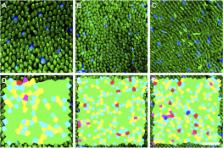
          Ground squirrels are an increasingly important model for studying visual processing, retinal circuitry, and cone photoreceptor function. Here, we demonstrate that the photoreceptor mosaic can be longitudinally imaged noninvasively in the 13-lined ground squirrel ( Ictidomys tridecemlineatus) using confocal and nonconfocal split-detection adaptive optics scanning ophthalmoscopy using 790 nm light. Photoreceptor density, spacing, and Voronoi analysis are consistent with that of the human cone mosaic. The high imaging success rate and consistent image quality in this study reinforce the ground squirrel as a practical model to aid drug discovery and testing through longitudinal imaging on the cellular scale.

          Related collections

          Most cited references43

          • Record: found
          • Abstract: found
          • Article: not found

          Adaptive optics scanning laser ophthalmoscopy.

          We present the first scanning laser ophthalmoscope that uses adaptive optics to measure and correct the high order aberrations of the human eye. Adaptive optics increases both lateral and axial resolution, permitting axial sectioning of retinal tissue in vivo. The instrument is used to visualize photoreceptors, nerve fibers and flow of white blood cells in retinal capillaries.
            Bookmark
            • Record: found
            • Abstract: found
            • Article: found
            Is Open Access

            Reflective afocal broadband adaptive optics scanning ophthalmoscope

            A broadband adaptive optics scanning ophthalmoscope (BAOSO) consisting of four afocal telescopes, formed by pairs of off-axis spherical mirrors in a non-planar arrangement, is presented. The non-planar folding of the telescopes is used to simultaneously reduce pupil and image plane astigmatism. The former improves the adaptive optics performance by reducing the root-mean-square (RMS) of the wavefront and the beam wandering due to optical scanning. The latter provides diffraction limited performance over a 3 diopter (D) vergence range. This vergence range allows for the use of any broadband light source(s) in the 450-850 nm wavelength range to simultaneously image any combination of retinal layers. Imaging modalities that could benefit from such a large vergence range are optical coherence tomography (OCT), multi- and hyper-spectral imaging, single- and multi-photon fluorescence. The benefits of the non-planar telescopes in the BAOSO are illustrated by resolving the human foveal photoreceptor mosaic in reflectance using two different superluminescent diodes with 680 and 796 nm peak wavelengths, reaching the eye with a vergence of 0.76 D relative to each other.
              Bookmark
              • Record: found
              • Abstract: found
              • Article: not found

              Intersubject variability of foveal cone photoreceptor density in relation to eye length.

              Adaptive optics scanning laser ophthalmoscopy (AOSLO) under optimized wavefront correction allows for routine imaging of foveal cone photoreceptors. The intersubject variability of foveal cone density was measured and its relation to eye length evaluated. AOSLO was used to image 18 healthy eyes with axial lengths from 22.86 to 28.31 mm. Ocular biometry and an eye model were used to estimate the retinal magnification factor. Individual cones in the AOSLO images were labeled, and the locations were used to generate topographic maps representing the spatial distribution of density. Representative retinal (cones/mm(2)) and angular (cones/deg(2)) cone densities at specific eccentricities were calculated from these maps. The entire foveal cone mosaic was resolved in four eyes, whereas the cones within 0.03 mm eccentricity remained unresolved in most eyes. The preferred retinal locus deviated significantly (P < 0.001) from the point of peak cone density for all except one individual. A significant decrease in retinal density (P < 0.05) with increasing axial length was observed at 0.30 mm eccentricity but not closer. Longer, more myopic eyes generally had higher angular density near the foveal center than the shorter eyes, but by 1°, this difference was nullified by retinal expansion, and so angular densities across all eyes were similar. The AOSLO can resolve the smallest foveal cones in certain eyes. Although myopia causes retinal stretching in the fovea, its effect within the foveola is confounded by factors other than cone density that have high levels of intersubject variability.
                Bookmark

                Author and article information

                Journal
                8809466
                1857
                Vis Neurosci
                Vis. Neurosci.
                Visual neuroscience
                0952-5238
                1469-8714
                11 March 2016
                January 2016
                16 March 2016
                : 33
                : E003
                Affiliations
                [1 ]Department of Cell Biology, Neurobiology, & Anatomy, Medical College of Wisconsin, Milwaukee, Wisconsin
                [2 ]Department of Ophthalmology, Medical College of Wisconsin, Milwaukee, Wisconsin
                [3 ]Neuroscience Research Institute, University of California, Santa Barbara, Santa Barbara, California
                [4 ]Department of Biology, University of Wisconsin Oshkosh, Oshkosh, Wisconsin
                [5 ]Department of Biomedical Engineering, Marquette University, Milwaukee, Wisconsin
                Author notes
                Address correspondence to: Alfredo Dubra, Medical College of Wisconsin, The Eye Institute, 925 N. 87th Street, Milwaukee, WI 53226. adubra@ 123456mcw.edu
                Article
                NIHMS766769
                10.1017/S0952523815000346
                4793898
                26923645
                f9c42b74-6068-43b7-85a3-73df15151064

                This is an Open Access article, distributed under the terms of the Creative Commons Attribution licence ( http://creativecommons.org/licenses/by/4.0/), which permits unrestricted re-use, distribution, and reproduction in any medium, provided the original work is properly cited.

                History
                Categories
                Article

                photoreceptors,adaptive optics,visual streak,rodent,retinal imaging

                Comments

                Comment on this article