39
views
0
recommends
+1 Recommend
0 collections
    0
    shares
      • Record: found
      • Abstract: found
      • Article: found
      Is Open Access

      Role of putative voltage-sensor countercharge D4 in regulating gating properties of Ca V1.2 and Ca V1.3 calcium channels

      research-article

      Read this article at

      Bookmark
          There is no author summary for this article yet. Authors can add summaries to their articles on ScienceOpen to make them more accessible to a non-specialist audience.

          ABSTRACT

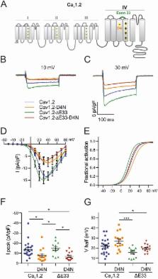
          Voltage-dependent calcium channels (Ca V) activate over a wide range of membrane potentials, and the voltage-dependence of activation of specific channel isoforms is exquisitely tuned to their diverse functions in excitable cells. Alternative splicing further adds to the stunning diversity of gating properties. For example, developmentally regulated insertion of an alternatively spliced exon 29 in the fourth voltage-sensing domain (VSD IV) of Ca V1.1 right-shifts voltage-dependence of activation by 30 mV and decreases the current amplitude several-fold. Previously we demonstrated that this regulation of gating properties depends on interactions between positive gating charges (R1, R2) and a negative countercharge (D4) in VSD IV of Ca V1.1. Here we investigated whether this molecular mechanism plays a similar role in the VSD IV of Ca V1.3 and in VSDs II and IV of Ca V1.2 by introducing charge-neutralizing mutations (D4N or E4Q) in the corresponding positions of Ca V1.3 and in two splice variants of Ca V1.2. In both channels the D4N (VSD IV) mutation resulted in a  ̴5 mV right-shift of the voltage-dependence of activation and in a reduction of current density to about half of that in controls. However in Ca V1.2 the effects were independent of alternative splicing, indicating that the two modulatory processes operate by distinct mechanisms. Together with our previous findings these results suggest that molecular interactions engaging D4 in VSD IV contribute to voltage-sensing in all examined Ca V1 channels, however its striking role in regulating the gating properties by alternative splicing appears to be a unique property of the skeletal muscle Ca V1.1 channel.

          Related collections

          Most cited references30

          • Record: found
          • Abstract: found
          • Article: not found

          Voltage-gated calcium channels.

          Voltage-gated calcium (Ca(2+)) channels are key transducers of membrane potential changes into intracellular Ca(2+) transients that initiate many physiological events. There are ten members of the voltage-gated Ca(2+) channel family in mammals, and they serve distinct roles in cellular signal transduction. The Ca(V)1 subfamily initiates contraction, secretion, regulation of gene expression, integration of synaptic input in neurons, and synaptic transmission at ribbon synapses in specialized sensory cells. The Ca(V)2 subfamily is primarily responsible for initiation of synaptic transmission at fast synapses. The Ca(V)3 subfamily is important for repetitive firing of action potentials in rhythmically firing cells such as cardiac myocytes and thalamic neurons. This article presents the molecular relationships and physiological functions of these Ca(2+) channel proteins and provides information on their molecular, genetic, physiological, and pharmacological properties.
            Bookmark
            • Record: found
            • Abstract: found
            • Article: not found

            Structure of the voltage-gated calcium channel Cav1.1 at 3.6 Å resolution.

            The voltage-gated calcium (Cav) channels convert membrane electrical signals to intracellular Ca(2+)-mediated events. Among the ten subtypes of Cav channel in mammals, Cav1.1 is specified for the excitation-contraction coupling of skeletal muscles. Here we present the cryo-electron microscopy structure of the rabbit Cav1.1 complex at a nominal resolution of 3.6 Å. The inner gate of the ion-conducting α1-subunit is closed and all four voltage-sensing domains adopt an 'up' conformation, suggesting a potentially inactivated state. The extended extracellular loops of the pore domain, which are stabilized by multiple disulfide bonds, form a windowed dome above the selectivity filter. One side of the dome provides the docking site for the α2δ-1-subunit, while the other side may attract cations through its negative surface potential. The intracellular I-II and III-IV linker helices interact with the β1a-subunit and the carboxy-terminal domain of α1, respectively. Classification of the particles yielded two additional reconstructions that reveal pronounced displacement of β1a and adjacent elements in α1. The atomic model of the Cav1.1 complex establishes a foundation for mechanistic understanding of excitation-contraction coupling and provides a three-dimensional template for molecular interpretations of the functions and disease mechanisms of Cav and Nav channels.
              Bookmark
              • Record: found
              • Abstract: found
              • Article: found
              Is Open Access

              The hitchhiker’s guide to the voltage-gated sodium channel galaxy

              Eukaryotic voltage-gated sodium (Nav) channels contribute to the rising phase of action potentials and served as an early muse for biophysicists laying the foundation for our current understanding of electrical signaling. Given their central role in electrical excitability, it is not surprising that (a) inherited mutations in genes encoding for Nav channels and their accessory subunits have been linked to excitability disorders in brain, muscle, and heart; and (b) Nav channels are targeted by various drugs and naturally occurring toxins. Although the overall architecture and behavior of these channels are likely to be similar to the more well-studied voltage-gated potassium channels, eukaryotic Nav channels lack structural and functional symmetry, a notable difference that has implications for gating and selectivity. Activation of voltage-sensing modules of the first three domains in Nav channels is sufficient to open the channel pore, whereas movement of the domain IV voltage sensor is correlated with inactivation. Also, structure–function studies of eukaryotic Nav channels show that a set of amino acids in the selectivity filter, referred to as DEKA locus, is essential for Na+ selectivity. Structures of prokaryotic Nav channels have also shed new light on mechanisms of drug block. These structures exhibit lateral fenestrations that are large enough to allow drugs or lipophilic molecules to gain access into the inner vestibule, suggesting that this might be the passage for drug entry into a closed channel. In this Review, we will synthesize our current understanding of Nav channel gating mechanisms, ion selectivity and permeation, and modulation by therapeutics and toxins in light of the new structures of the prokaryotic Nav channels that, for the time being, serve as structural models of their eukaryotic counterparts.
                Bookmark

                Author and article information

                Journal
                Channels (Austin)
                Channels (Austin)
                KCHL
                kchl20
                Channels
                Taylor & Francis
                1933-6950
                1933-6969
                2018
                26 September 2018
                26 September 2018
                : 12
                : 1
                : 249-261
                Affiliations
                [a ]Department of Physiology and Medical Physics, Medical University of Innsbruck , Innsbruck, Austria
                [b ]Institute of Experimental Neuroregeneration Spinal Cord Injury and Tissue Regeneration Center Salzburg (SCI-TReCS), Paracelsus Medical University , Salzburg, Austria
                [c ]Department of Pharmacology and Toxicology, University of Innsbruck , Innsbruck, Austria
                Author notes
                CONTACT Bernhard E. Flucher bernhard.e.flucher@ 123456i-med.ac.at
                Author information
                http://orcid.org/0000-0002-3583-9223
                http://orcid.org/0000-0002-9629-2073
                http://orcid.org/0000-0002-4286-5067
                http://orcid.org/0000-0003-3660-6138
                http://orcid.org/0000-0002-5255-4705
                Article
                1482183
                10.1080/19336950.2018.1482183
                6161609
                30001160
                f99cb702-e34e-401c-ae77-b33630ce3e04
                © 2018 The Author(s). Published by Informa UK Limited, trading as Taylor & Francis Group.

                This is an Open Access article distributed under the terms of the Creative Commons Attribution License ( http://creativecommons.org/licenses/by/4.0/), which permits unrestricted use, distribution, and reproduction in any medium, provided the original work is properly cited.

                History
                : 17 May 2018
                : 22 May 2018
                Page count
                Figures: 5, Tables: 3, References: 34, Pages: 13
                Funding
                Funded by: Austrian Science Fund 10.13039/501100002428
                Award ID: P27031
                Award ID: P30402
                Award ID: F4406
                Award ID: W1101
                Funded by: University of Innsbruck
                Award ID: P7400-027-011; P7400-027-012
                This work was supported by the Austrian Science Fund [P27031, P30402, F4406, W1101]; University of Innsbruck [P7400-027-011; P7400-027-012].
                Categories
                Research Paper

                Molecular biology
                alternative splicing,voltage gated calcium channels,voltage-sensing
                Molecular biology
                alternative splicing, voltage gated calcium channels, voltage-sensing

                Comments

                Comment on this article