13
views
0
recommends
+1 Recommend
0 collections
    0
    shares
      • Record: found
      • Abstract: found
      • Article: found
      Is Open Access

      Phenotypic screen for oxygen consumption rate identifies an anti-cancer naphthoquinone that induces mitochondrial oxidative stress

      research-article

      Read this article at

      Bookmark
          There is no author summary for this article yet. Authors can add summaries to their articles on ScienceOpen to make them more accessible to a non-specialist audience.

          Abstract

          A hallmark of cancer cells is their ability to reprogram nutrient metabolism. Thus, disruption to this phenotype is a potential avenue for anti-cancer therapy. Herein we used a phenotypic chemical library screening approach to identify molecules that disrupted nutrient metabolism (by increasing cellular oxygen consumption rate) and were toxic to cancer cells. From this screen we discovered a 1,4-Naphthoquinone (referred to as BH10) that is toxic to a broad range of cancer cell types. BH10 has improved cancer-selective toxicity compared to doxorubicin, 17-AAG, vitamin K3, and other known anti-cancer quinones. BH10 increases glucose oxidation via both mitochondrial and pentose phosphate pathways, decreases glycolysis, lowers GSH:GSSG and NAPDH/NAPD + ratios exclusively in cancer cells, and induces necrosis. BH10 targets mitochondrial redox defence as evidenced by increased mitochondrial peroxiredoxin 3 oxidation and decreased mitochondrial aconitase activity, without changes in markers of cytosolic or nuclear damage. Over-expression of mitochondria-targeted catalase protects cells from BH10-mediated toxicity, while the thioredoxin reductase inhibitor auranofin synergistically enhances BH10-induced peroxiredoxin 3 oxidation and cytotoxicity. Overall, BH10 represents a 1,4-Naphthoquinone with an improved cancer-selective cytotoxicity profile via its mitochondrial specificity.
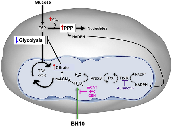
          Graphical abstract

          Schematic overview of BH10-induced effects on cancer cells. Treatment of cancer cells with BH10 induced mitochondrial oxidative stress. Activity of the redox-sensitive enzyme mitochondrial aconitase (mACN) was impaired leading to citrate accumulation and a decrease in glycolysis. BH10 increased pentose phosphate pathway activity to increase NADPH production for antioxidant defence. Mitochondrial oxidative stress is evidenced by oxidation of the peroxiredoxin/thioredoxin reductase system, including peroxiredoxin 3 dimerization (oxidation), and depletion of NADPH. BH10's cytotoxicity and effect on PRDX3 dimerization were synergistically enhanced with the thioredoxin reductase inhibitor auranofin, while over-expression of mitochondrial-targeted catalase (mCAT) or treatment with antioxidants N-acetylcysteine (NAC) and GSH protected cells from BH10-induced death.

          Highlights

          • Phenotypic screen identifies a 1,4-Naphthoquinone BH10 with improved cancer-selective cytotoxicity

          • BH10 decreases glycolysis and increases glucose oxidation

          • BH10-mediated cytotoxicity is linked to mitochondrial oxidative stress

          Related collections

          Most cited references22

          • Record: found
          • Abstract: found
          • Article: found
          Is Open Access

          Glutathione metabolism in cancer progression and treatment resistance

          Bansal and Simon discuss strategies to block glutathione synthesis and utilization pathways to inhibit tumor propagation and treatment resistance.
            Bookmark
            • Record: found
            • Abstract: found
            • Article: not found

            Dichloroacetate (DCA) as a potential metabolic-targeting therapy for cancer

            The unique metabolism of most solid tumours (aerobic glycolysis, i.e., Warburg effect) is not only the basis of diagnosing cancer with metabolic imaging but might also be associated with the resistance to apoptosis that characterises cancer. The glycolytic phenotype in cancer appears to be the common denominator of diverse molecular abnormalities in cancer and may be associated with a (potentially reversible) suppression of mitochondrial function. The generic drug dichloroacetate is an orally available small molecule that, by inhibiting the pyruvate dehydrogenase kinase, increases the flux of pyruvate into the mitochondria, promoting glucose oxidation over glycolysis. This reverses the suppressed mitochondrial apoptosis in cancer and results in suppression of tumour growth in vitro and in vivo. Here, we review the scientific and clinical rationale supporting the rapid translation of this promising metabolic modulator in early-phase cancer clinical trials.
              Bookmark
              • Record: found
              • Abstract: found
              • Article: not found

              Akt/protein kinase B is constitutively active in non-small cell lung cancer cells and promotes cellular survival and resistance to chemotherapy and radiation.

              To evaluate the role of Akt/PKB in non-small cell lung cancer (NSCLC) survival, we analyzed NSCLC cell lines that differed in tumor histology as well as p53, Rb, and K-ras status. Constitutive Akt/protein kinase B (PKB) activity was demonstrated in 16 of 17 cell lines by maintenance of S473 phosphorylation with serum deprivation. Additional analysis of five of 2these NSCLC lines revealed that phosphorylation of S473 and T308 correlated with in vitro kinase activity. Akt/PKB activation was phosphatidylinositol 3-kinase-dependent and promoted survival because the phosphatidylinositol 3 inhibitors LY294002 and wortmannin inhibited Akt/PKB phosphorylation, Akt/PKB activity, and increased apoptosis only in cells with active Akt/PKB. To test whether Akt/PKB activity promoted therapeutic resistance, LY294002 was added with individual chemotherapeutic agents or irradiation. LY294002 greatly potentiated chemotherapy-induced apoptosis in cells with high Akt/PKB levels, but did not significantly increase chemotherapy-induced apoptosis in cells with low Akt/PKB levels. Combined with radiation in cells with active Akt/PKB, LY294002 additively increased apoptosis and inhibited clonogenic growth. These results were extended with transiently transfected Akt/PKB mutants. Transfecting dominant negative Akt/PKB decreased Akt/PKB activity and increased basal apoptosis as well as chemotherapy- and irradiation-induced apoptosis only in cells with high Akt/PKB activity. Conversely, transfecting constitutively active Akt/PKB into cells with low Akt/PKB activity increased Akt/PKB activity and attenuated chemotherapy- and radiation-induced apoptosis. We therefore identify Akt/PKB as a constitutively active kinase that promotes survival of NSCLC cells and demonstrate that modulation of Akt/PKB activity by pharmacological or genetic approaches alters the cellular responsiveness to therapeutic modalities typically used to treat patients with NSCLC.
                Bookmark

                Author and article information

                Contributors
                Journal
                Redox Biol
                Redox Biol
                Redox Biology
                Elsevier
                2213-2317
                05 November 2019
                January 2020
                05 November 2019
                : 28
                : 101374
                Affiliations
                [a ]School of Biotechnology and Biomolecular Sciences, University of New South Wales, Sydney, NSW, Australia
                [b ]School of Mathematics and Statistics, The University of Sydney, Sydney, Australia
                [c ]School of Chemistry, University of New South Wales, Sydney, NSW, Australia
                [d ]Charles Perkins Centre, School of Life and Environmental Sciences, University of Sydney, Australia
                [e ]Department of Chemistry and VT Center for Drug Discovery, Virginia Tech, Blacksburg, VA, USA
                [f ]Department of Biochemistry and Genetics, La Trobe Institute for Molecular Science, La Trobe University, Melbourne, VIC, Australia
                [g ]Walter and Eliza Hall Institute of Medical Research, Melbourne, VIC, Australia
                [h ]Concord Medical School, Asbestos Disease Research Institute, University of Sydney, Australia
                [i ]Children's Cancer Institute, Lowy Cancer Research Centre, University of New South Wales Sydney, New South Wales, Australia
                [j ]School of Medical Sciences, University of New South Wales, Sydney, NSW, Australia
                Author notes
                []Corresponding author.School of Biotechnology and Biomolecular Sciences, Level 4 D26 Biological Sciences Building, University of New South Wales, Sydney, 2052, Australia. frances.byrne@ 123456unsw.edu.au
                [∗∗ ]Corresponding author. k.hoehn@ 123456unsw.edu.au
                Article
                S2213-2317(19)30884-5 101374
                10.1016/j.redox.2019.101374
                6861633
                31743887
                f84e4ee8-7240-4e6d-965f-100c2b2823cf
                © 2019 The Authors

                This is an open access article under the CC BY-NC-ND license (http://creativecommons.org/licenses/by-nc-nd/4.0/).

                History
                : 31 July 2019
                : 7 October 2019
                : 4 November 2019
                Categories
                Research Paper

                cancer metabolism,quinone,peroxiredoxin,mitochondria
                cancer metabolism, quinone, peroxiredoxin, mitochondria

                Comments

                Comment on this article