14
views
0
recommends
+1 Recommend
0 collections
    0
    shares
      • Record: found
      • Abstract: found
      • Article: found
      Is Open Access

      Obesity-induced inflammation: The impact of the hematopoietic stem cell niche

      review-article
      ,
      JCI Insight
      American Society for Clinical Investigation

      Read this article at

      Bookmark
          There is no author summary for this article yet. Authors can add summaries to their articles on ScienceOpen to make them more accessible to a non-specialist audience.

          Abstract

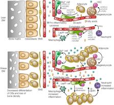
          Obesity and obesity-related diseases like type 2 diabetes (T2D) are prominent global health issues; therefore, there is a need to better understand the mechanisms underlying these conditions. The onset of obesity is characterized by accumulation of proinflammatory cells, including Ly6c hi monocytes (which differentiate into proinflammatory macrophages) and neutrophils, in metabolic tissues. This shift toward chronic, low-grade inflammation is an obese-state hallmark and highly linked to metabolic disorders and other obesity comorbidities. The mechanisms that induce and maintain increased inflammatory myelopoiesis are of great interest, with a recent focus on how obesity affects more primitive hematopoietic cells. The hematopoietic system is constantly replenished by proper regulation of hematopoietic stem and progenitor (HSPC) pools in the BM. While early research suggests that chronic obesity promotes expansion of myeloid-skewed HSPCs, the involvement of the hematopoietic stem cell (HSC) niche in regulating obesity-induced myelopoiesis remains undefined. In this review, we explore the role of the multicellular HSC niche in hematopoiesis and inflammation, and the potential contribution of this niche to the hematopoietic response to obesity. This review further aims to summarize the potential HSC niche involvement as a target of obesity-induced inflammation and a driver of obesity-induced myelopoiesis.

          Related collections

          Most cited references199

          • Record: found
          • Abstract: found
          • Article: not found

          MCP-1 contributes to macrophage infiltration into adipose tissue, insulin resistance, and hepatic steatosis in obesity.

          Adipocytes secrete a variety of bioactive molecules that affect the insulin sensitivity of other tissues. We now show that the abundance of monocyte chemoattractant protein-1 (MCP-1) mRNA in adipose tissue and the plasma concentration of MCP-1 were increased both in genetically obese diabetic (db/db) mice and in WT mice with obesity induced by a high-fat diet. Mice engineered to express an MCP-1 transgene in adipose tissue under the control of the aP2 gene promoter exhibited insulin resistance, macrophage infiltration into adipose tissue, and increased hepatic triglyceride content. Furthermore, insulin resistance, hepatic steatosis, and macrophage accumulation in adipose tissue induced by a high-fat diet were reduced extensively in MCP-1 homozygous KO mice compared with WT animals. Finally, acute expression of a dominant-negative mutant of MCP-1 ameliorated insulin resistance in db/db mice and in WT mice fed a high-fat diet. These findings suggest that an increase in MCP-1 expression in adipose tissue contributes to the macrophage infiltration into this tissue, insulin resistance, and hepatic steatosis associated with obesity in mice.
            Bookmark
            • Record: found
            • Abstract: found
            • Article: not found

            Obesity induces a phenotypic switch in adipose tissue macrophage polarization.

            Adipose tissue macrophages (ATMs) infiltrate adipose tissue during obesity and contribute to insulin resistance. We hypothesized that macrophages migrating to adipose tissue upon high-fat feeding may differ from those that reside there under normal diet conditions. To this end, we found a novel F4/80(+)CD11c(+) population of ATMs in adipose tissue of obese mice that was not seen in lean mice. ATMs from lean mice expressed many genes characteristic of M2 or "alternatively activated" macrophages, including Ym1, arginase 1, and Il10. Diet-induced obesity decreased expression of these genes in ATMs while increasing expression of genes such as those encoding TNF-alpha and iNOS that are characteristic of M1 or "classically activated" macrophages. Interestingly, ATMs from obese C-C motif chemokine receptor 2-KO (Ccr2-KO) mice express M2 markers at levels similar to those from lean mice. The antiinflammatory cytokine IL-10, which was overexpressed in ATMs from lean mice, protected adipocytes from TNF-alpha-induced insulin resistance. Thus, diet-induced obesity leads to a shift in the activation state of ATMs from an M2-polarized state in lean animals that may protect adipocytes from inflammation to an M1 proinflammatory state that contributes to insulin resistance.
              Bookmark
              • Record: found
              • Abstract: found
              • Article: not found

              Mesenchymal and haematopoietic stem cells form a unique bone marrow niche.

              The cellular constituents forming the haematopoietic stem cell (HSC) niche in the bone marrow are unclear, with studies implicating osteoblasts, endothelial and perivascular cells. Here we demonstrate that mesenchymal stem cells (MSCs), identified using nestin expression, constitute an essential HSC niche component. Nestin(+) MSCs contain all the bone-marrow colony-forming-unit fibroblastic activity and can be propagated as non-adherent 'mesenspheres' that can self-renew and expand in serial transplantations. Nestin(+) MSCs are spatially associated with HSCs and adrenergic nerve fibres, and highly express HSC maintenance genes. These genes, and others triggering osteoblastic differentiation, are selectively downregulated during enforced HSC mobilization or beta3 adrenoreceptor activation. Whereas parathormone administration doubles the number of bone marrow nestin(+) cells and favours their osteoblastic differentiation, in vivo nestin(+) cell depletion rapidly reduces HSC content in the bone marrow. Purified HSCs home near nestin(+) MSCs in the bone marrow of lethally irradiated mice, whereas in vivo nestin(+) cell depletion significantly reduces bone marrow homing of haematopoietic progenitors. These results uncover an unprecedented partnership between two distinct somatic stem-cell types and are indicative of a unique niche in the bone marrow made of heterotypic stem-cell pairs.
                Bookmark

                Author and article information

                Contributors
                Journal
                JCI Insight
                JCI Insight
                JCI Insight
                JCI Insight
                American Society for Clinical Investigation
                2379-3708
                8 February 2021
                8 February 2021
                8 February 2021
                : 6
                : 3
                : e145295
                Affiliations
                Department of Pediatrics and Department of Molecular and Integrative Physiology, University of Michigan Medical School, Ann Arbor, Michigan, USA.
                Author notes
                Address correspondence to: Kanakadurga Singer, Department of Pediatrics, Division of Pediatric Endocrinology, D1205 MPB,1500 E Medical Center Dr., Ann Arbor, Michigan 48109, USA. Phone: 734.764.5175; Email: ksinger@ 123456umich.edu .
                Author information
                http://orcid.org/0000-0001-8278-3800
                Article
                145295
                10.1172/jci.insight.145295
                7934850
                33554957
                f8456301-f68c-4c60-a548-4bea74224c59
                © 2021 Bowers et al.

                This work is licensed under the Creative Commons Attribution 4.0 International License. To view a copy of this license, visit http://creativecommons.org/licenses/by/4.0/.

                History
                Categories
                Review

                Comments

                Comment on this article