36
views
0
recommends
+1 Recommend
0 collections
    0
    shares
      • Record: found
      • Abstract: found
      • Article: found
      Is Open Access

      Impaired Bone Regenerative Effect of Exosomes Derived from Bone Marrow Mesenchymal Stem Cells in Type 1 Diabetes

      research-article

      Read this article at

      Bookmark
          There is no author summary for this article yet. Authors can add summaries to their articles on ScienceOpen to make them more accessible to a non-specialist audience.

          Abstract

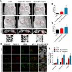
          Stem cell‐derived exosomes have exhibited promise for applications in tissue regeneration. However, one major problem for stem cell‐derived exosome therapies is identifying appropriate source cells. In the present study, we aimed to compare the bone regenerative effect of exosomes secreted by bone marrow mesenchymal stem cells (BMSCs) derived from type 1 diabetes rats (dBMSC‐exos) and exosomes secreted by BMSCs derived from normal rats (nBMSC‐exos). BMSCs were isolated from rats with streptozotocin‐induced diabetes and normal rats. dBMSC‐exos and nBMSC‐exos were isolated by an ultracentrifugation method and identified. The effects of dBMSC‐exos and nBMSC‐exos on the proliferation and migration of BMSCs and human umbilical vein endothelial cells (HUVECs) were investigated. The effects of exosomes on the osteogenic differentiation of BMSCs and the angiogenic activity of HUVECs were compared. Finally, a rat calvarial defect model was used to compare the effects of exosomes on bone regeneration and neovascularization in vivo. In vitro, dBMSC‐exos and nBMSC‐exos both enhanced the osteogenic differentiation of BMSCs and promoted the angiogenic activity of HUVECs, but nBMSC‐exos had a greater effect than dBMSC‐exos. Similarly, in vivo, both dBMSC‐exos and nBMSC‐exos promoted bone regeneration and neovascularization in rat calvarial defects, but the therapeutic effect of nBMSC‐exos was superior to that of dBMSC‐exos. The present study demonstrates for the first time that the bone regenerative effect of exosomes derived from BMSCs is impaired in type 1 diabetes, indicating that for patients with type 1 diabetes, the autologous transplantation of BMSC‐exos to promote bone regeneration may be inappropriate. stem cells translational medicine 2019;8:593–605

          Related collections

          Most cited references38

          • Record: found
          • Abstract: found
          • Article: found
          Is Open Access

          Exosomes from human umbilical cord blood accelerate cutaneous wound healing through miR-21-3p-mediated promotion of angiogenesis and fibroblast function

          The application of blood plasma for soft tissue wound healing is receiving much more attention recently. Exosomes are critical paracrine mediators that can be obtained from biological fluids including plasma and be able to induce regenerative effects by transferring bioactive molecules such as microRNAs (miRNAs). This study aimed to investigate the effects of exosomes from human umbilical cord blood plasma (UCB-Exos) on wound healing and to elucidate the underlying mechanism. Methods: UCB-Exos were isolated by ultracentrifugation and subcutaneously injected into full-thickness skin wounds in mice. The efficacy of UCB-Exos on wound healing was evaluated by measuring wound closure rates, histological analysis and immunofluorescence examinations. In vitro, quantitative real-time PCR (qRT-PCR) analysis was performed to detect the expression levels of a class of miRNAs that have positive roles in regulating wound healing. The scratch wound assay, transwell assay and cell counting kit-8 analysis were conducted to assess the effects of UCB-Exos on migration and proliferation of human skin fibroblasts and endothelial cells. Tube formation assay was carried out to test the impact of UCB-Exos on angiogenic tube formation ability of endothelial cells. Meanwhile, by using specific RNA inhibitors or siRNAs, the roles of the candidate miRNA and its target genes in UCB-Exos-induced regulation of function of fibroblasts and endothelial cells were assessed. Results: The local transplantation of UCB-Exos into mouse skin wounds resulted in accelerated re-epithelialization, reduced scar widths, and enhanced angiogenesis. In vitro, UCB-Exos could promote the proliferation and migration of fibroblasts, and enhance the angiogenic activities of endothelial cells. Notably, miR-21-3p was found to be highly enriched in UCB-Exos and served as a critical mediator in UCB-Exos -induced regulatory effects through inhibition of phosphatase and tensin homolog (PTEN) and sprouty homolog 1 (SPRY1). Conclusion: Our results suggest that UCB-Exos are important effectors of plasma activity and can be used as a novel promising strategy for soft tissue wound healing.
            Bookmark
            • Record: found
            • Abstract: found
            • Article: found
            Is Open Access

            Exosomes/tricalcium phosphate combination scaffolds can enhance bone regeneration by activating the PI3K/Akt signaling pathway

            Background Recently, accumulating evidence has shown that exosomes, the naturally secreted nanocarriers of cells, can exert therapeutic effects in various disease models in the absence of parent cells. However, application of exosomes in bone defect repair and regeneration has been rarely reported, and little is known regarding their underlying mechanisms. Methods Exosomes derived from human-induced pluripotent stem cell-derived mesenchymal stem cells (hiPS-MSC-Exos) were combined with tricalcium phosphate (β-TCP) to repair critical-sized calvarial bone defects, and the efficacy was assessed by histological examination. We evaluated the in vitro effects of hiPSC-MSC-Exos on the proliferation, migration, and osteogenic differentiation of human bone marrow-derived mesenchymal stem cells (hBMSCs) by cell-counting, scratch assays, and qRT-PCR, respectively. Gene expression profiling and bioinformatics analyses were also used to identify the underlying mechanisms in the repair. Results We found that the exosome/β-TCP combination scaffolds could enhance osteogenesis as compared to pure β-TCP scaffolds. In vitro assays showed that the exosomes could release from β-TCP and could be internalized by hBMSCs. In addition, the internalization of exosomes into hBMSCs could profoundly enhance the proliferation, migration, and osteogenic differentiation of hBMSCs. Furthermore, gene expression profiling and bioinformatics analyses demonstrated that exosome/β-TCP combination scaffolds significantly altered the expression of a network of genes involved in the PI3K/Akt signaling pathway. Functional studies further confirmed that the PI3K/Akt signaling pathway was the critical mediator during the exosome-induced osteogenic responses of hBMSCs. Conclusions We propose that the exosomes can enhance the osteoinductivity of β-TCP through activating the PI3K/Akt signaling pathway of hBMSCs, which means that the exosome/β-TCP combination scaffolds possess better osteogenesis activity than pure β-TCP scaffolds. These results indicate that naturally secreted nanocarriers-exosomes can be used as a bioactive material to improve the bioactivity of the biomaterials, and that hiPS-MSC-Exos combined with β-TCP scaffolds can be potentially used for repairing bone defects. Electronic supplementary material The online version of this article (doi:10.1186/s13287-016-0391-3) contains supplementary material, which is available to authorized users.
              Bookmark
              • Record: found
              • Abstract: found
              • Article: found
              Is Open Access

              The role of vasculature in bone development, regeneration and proper systemic functioning

              Bone is a richly vascularized connective tissue. As the main source of oxygen, nutrients, hormones, neurotransmitters and growth factors delivered to the bone cells, vasculature is indispensable for appropriate bone development, regeneration and remodeling. Bone vasculature also orchestrates the process of hematopoiesis. Blood supply to the skeletal system is provided by the networks of arteries and arterioles, having distinct molecular characteristics and localizations within the bone structures. Blood vessels of the bone develop through the process of angiogenesis, taking place through different, bone-specific mechanisms. Impaired functioning of the bone blood vessels may be associated with the occurrence of some skeletal and systemic diseases, i.e., osteonecrosis, osteoporosis, atherosclerosis or diabetes mellitus. When a disease or trauma-related large bone defects appear, bone grafting or bone tissue engineering-based strategies are required. However, a successful bone regeneration in both approaches largely depends on a proper blood supply. In this paper, we review the most recent data on the functions, molecular characteristics and significance of the bone blood vessels, with a particular emphasis on the role of angiogenesis and blood vessel functioning in bone development and regeneration, as well as the consequences of its impairment in the course of different skeletal and systemic diseases.
                Bookmark

                Author and article information

                Contributors
                xujia0117@126.com
                ymchai@sjtu.edu.cn
                Journal
                Stem Cells Transl Med
                Stem Cells Transl Med
                10.1002/(ISSN)2157-6580
                SCT3
                Stem Cells Translational Medicine
                John Wiley & Sons, Inc. (Hoboken, USA )
                2157-6564
                2157-6580
                26 February 2019
                June 2019
                : 8
                : 6 ( doiID: 10.1002/sct3.2019.8.issue-6 )
                : 593-605
                Affiliations
                [ 1 ] Department of Orthopedic Surgery Shanghai Jiao Tong University Affiliated Sixth People's Hospital Shanghai People's Republic of China
                Author notes
                [*] [* ]Correspondence: Yimin Chai, M.D., Ph.D., Department of Orthopedic Surgery, Shanghai Jiao Tong University Affiliated Sixth People's Hospital, Yishan Rd. 600, Shanghai 200233, People's Republic of China. Telephone: 0086‐21‐64369181; e‐mail: ymchai@ 123456sjtu.edu.cn ; or Jia Xu, M.D., Ph.D., Department of Orthopedic Surgery, Shanghai Jiao Tong University Affiliated Sixth People's Hospital, Yishan Rd. 600, Shanghai 200233, People's Republic of China. Telephone: 0086‐13611960282; e‐mail: xujia0117@ 123456126.com
                [†]

                Contributed equally.

                Author information
                https://orcid.org/0000-0002-1972-5971
                Article
                SCT312468
                10.1002/sctm.18-0199
                6525563
                30806487
                f7825b58-1154-4044-923a-5b8af19a5a3e
                © 2019 The Authors. stem cells translational medicine published by Wiley Periodicals, Inc. on behalf of AlphaMed Press

                This is an open access article under the terms of the http://creativecommons.org/licenses/by-nc-nd/4.0/ License, which permits use and distribution in any medium, provided the original work is properly cited, the use is non‐commercial and no modifications or adaptations are made.

                History
                : 13 September 2018
                : 16 January 2019
                Page count
                Figures: 8, Tables: 0, Pages: 14, Words: 8778
                Funding
                Funded by: National Natural Science Foundation of China
                Award ID: 81572122
                Award ID: 81772338
                Funded by: Interdisciplinary Program of Shanghai Jiao Tong University
                Award ID: YG2017ZD05
                Funded by: Shanghai Leading Talent Plan
                Award ID: 043
                Categories
                Tissue Engineering and Regenerative Medicine
                Tissue Engineering and Regenerative Medicine
                Custom metadata
                2.0
                sct312468
                June 2019
                Converter:WILEY_ML3GV2_TO_NLMPMC version:5.6.3 mode:remove_FC converted:18.05.2019

                exosomes,type 1 diabetes,osteogenesis,angiogenesis,bone regeneration

                Comments

                Comment on this article

                scite_
                0
                0
                0
                0
                Smart Citations
                0
                0
                0
                0
                Citing PublicationsSupportingMentioningContrasting
                View Citations

                See how this article has been cited at scite.ai

                scite shows how a scientific paper has been cited by providing the context of the citation, a classification describing whether it supports, mentions, or contrasts the cited claim, and a label indicating in which section the citation was made.

                Similar content326

                Cited by48

                Most referenced authors441