48
views
0
recommends
+1 Recommend
0 collections
    0
    shares
      • Record: found
      • Abstract: found
      • Article: found
      Is Open Access

      Emotion Reactivity Is Increased 4-6 Weeks Postpartum in Healthy Women: A Longitudinal fMRI Study

      research-article

      Read this article at

      Bookmark
          There is no author summary for this article yet. Authors can add summaries to their articles on ScienceOpen to make them more accessible to a non-specialist audience.

          Abstract

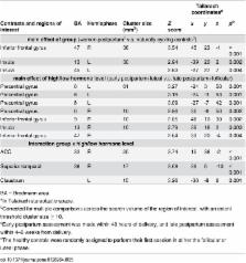
          Marked endocrine alterations occur after delivery. Most women cope well with these changes, but the postpartum period is associated with an increased risk of depressive episodes. Previous studies of emotion processing have focused on maternal–infant bonding or postpartum depression (PPD), and longitudinal studies of the neural correlates of emotion processing throughout the postpartum period in healthy women are lacking. In this study, 13 women, without signs of post partum depression, underwent fMRI with an emotional face matching task and completed the MADRS-S, STAI-S, and EPDS within 48 h (early postpartum) and 4–6 weeks after delivery (late postpartum). Also, data from a previous study including 15 naturally cycling controls assessed in the luteal and follicular phase of the menstrual cycle was used. Women had lower reactivity in insula, middle frontal gyrus (MFG), and inferior frontal gyrus (IFG) in the early as compared to the late postpartum assessment. Insular reactivity was positively correlated with anxiety in the early postpartum period and with depressive symptoms late postpartum. Reactivity in insula and IFG were greater in postpartum women than in non-pregnant control subjects. Brain reactivity was not correlated with serum estradiol or progesterone levels. Increased reactivity in the insula, IFG, and MFG may reflect normal postpartum adaptation, but correlation with self-rated symptoms of depression and anxiety in these otherwise healthy postpartum women, may also suggest that these changes place susceptible women at increased risk of PPD. These findings contribute to our understanding of the neurobiological aspects of the postpartum period, which might shed light on the mechanisms underlying affective puerperal disorders, such as PPD.

          Related collections

          Most cited references51

          • Record: found
          • Abstract: found
          • Article: not found

          Detection of postnatal depression. Development of the 10-item Edinburgh Postnatal Depression Scale.

          The development of a 10-item self-report scale (EPDS) to screen for Postnatal Depression in the community is described. After extensive pilot interviews a validation study was carried out on 84 mothers using the Research Diagnostic Criteria for depressive illness obtained from Goldberg's Standardised Psychiatric Interview. The EPDS was found to have satisfactory sensitivity and specificity, and was also sensitive to change in the severity of depression over time. The scale can be completed in about 5 minutes and has a simple method of scoring. The use of the EPDS in the secondary prevention of Postnatal Depression is discussed.
            Bookmark
            • Record: found
            • Abstract: found
            • Article: not found

            Serotonin transporter genetic variation and the response of the human amygdala.

            A functional polymorphism in the promoter region of the human serotonin transporter gene (SLC6A4) has been associated with several dimensions of neuroticism and psychopathology, especially anxiety traits, but the predictive value of this genotype against these complex behaviors has been inconsistent. Serotonin [5- hydroxytryptamine, (5-HT)] function influences normal fear as well as pathological anxiety, behaviors critically dependent on the amygdala in animal models and in clinical studies. We now report that individuals with one or two copies of the short allele of the serotonin transporter (5-HTT) promoter polymorphism, which has been associated with reduced 5-HTT expression and function and increased fear and anxiety-related behaviors, exhibit greater amygdala neuronal activity, as assessed by BOLD functional magnetic resonance imaging, in response to fearful stimuli compared with individuals homozygous for the long allele. These results demonstrate genetically driven variation in the response of brain regions underlying human emotional behavior and suggest that differential excitability of the amygdala to emotional stimuli may contribute to the increased fear and anxiety typically associated with the short SLC6A4 allele.
              Bookmark
              • Record: found
              • Abstract: found
              • Article: not found

              Functional atlas of emotional faces processing: a voxel-based meta-analysis of 105 functional magnetic resonance imaging studies.

              Most of our social interactions involve perception of emotional information from the faces of other people. Furthermore, such emotional processes are thought to be aberrant in a range of clinical disorders, including psychosis and depression. However, the exact neurofunctional maps underlying emotional facial processing are not well defined. Two independent researchers conducted separate comprehensive PubMed (1990 to May 2008) searches to find all functional magnetic resonance imaging (fMRI) studies using a variant of the emotional faces paradigm in healthy participants. The search terms were: "fMRI AND happy faces," "fMRI AND sad faces," "fMRI AND fearful faces," "fMRI AND angry faces," "fMRI AND disgusted faces" and "fMRI AND neutral faces." We extracted spatial coordinates and inserted them in an electronic database. We performed activation likelihood estimation analysis for voxel-based meta-analyses. Of the originally identified studies, 105 met our inclusion criteria. The overall database consisted of 1785 brain coordinates that yielded an overall sample of 1600 healthy participants. Quantitative voxel-based meta-analysis of brain activation provided neurofunctional maps for 1) main effect of human faces; 2) main effect of emotional valence; and 3) modulatory effect of age, sex, explicit versus implicit processing and magnetic field strength. Processing of emotional faces was associated with increased activation in a number of visual, limbic, temporoparietal and prefrontal areas; the putamen; and the cerebellum. Happy, fearful and sad faces specifically activated the amygdala, whereas angry or disgusted faces had no effect on this brain region. Furthermore, amygdala sensitivity was greater for fearful than for happy or sad faces. Insular activation was selectively reported during processing of disgusted and angry faces. However, insular sensitivity was greater for disgusted than for angry faces. Conversely, neural response in the visual cortex and cerebellum was observable across all emotional conditions. Although the activation likelihood estimation approach is currently one of the most powerful and reliable meta-analytical methods in neuroimaging research, it is insensitive to effect sizes. Our study has detailed neurofunctional maps to use as normative references in future fMRI studies of emotional facial processing in psychiatric populations. We found selective differences between neural networks underlying the basic emotions in limbic and insular brain regions.
                Bookmark

                Author and article information

                Contributors
                Role: Academic Editor
                Journal
                PLoS One
                PLoS ONE
                plos
                plosone
                PLoS ONE
                Public Library of Science (San Francisco, CA USA )
                1932-6203
                10 June 2015
                2015
                : 10
                : 6
                : e0128964
                Affiliations
                [1 ]Department of Women’s and Children’s Health, Uppsala University, Uppsala, Sweden
                [2 ]Department of Psychology, Uppsala University, Uppsala, Sweden
                [3 ]Department of Radiology, Oncology and Radiation Science, Uppsala University, Uppsala, Sweden
                University of Rennes-1, FRANCE
                Author notes

                Competing Interests: Dr. Sundstrom-Poromaa serves occasionally on advisory boards or acts as invited speaker at scientific meetings for MSD, Bayer Health Care, and Lundbeck A/S. The competing interests of Dr. Sundström-Poromaa do not alter the authors’ adherence to PLOS ONE policies on sharing data and materials and these conflicts of interest have been clearly stated upon submission.

                Conceived and designed the experiments: MG ISP. Performed the experiments: MG EB HM JE SS AS KK JW ISP. Analyzed the data: MG EB HM ISP. Contributed reagents/materials/analysis tools: JW. Wrote the paper: MG EB HM JE SS AS KK JW ISP.

                Article
                PONE-D-14-40716
                10.1371/journal.pone.0128964
                4465482
                26061879
                f6ac84be-65aa-4163-b03f-c5e2bcb729c9
                Copyright @ 2015

                This is an open access article distributed under the terms of the Creative Commons Attribution License, which permits unrestricted use, distribution, and reproduction in any medium, provided the original author and source are credited

                History
                : 10 September 2014
                : 1 May 2015
                Page count
                Figures: 2, Tables: 5, Pages: 16
                Funding
                The major funding for the study comes from the Swedish Research Council project K2008-54X-200642-01-3 (Sundstrom Poromaa) and the Swedish Council for Working Life and Social Research projects 2007-1955 (Sundstrom Poromaa).
                Categories
                Research Article
                Custom metadata
                The data underlying the article are available at: http://datadryad.org/review?doi=doi:10.5061/dryad.r1pf4.

                Uncategorized
                Uncategorized

                Comments

                Comment on this article