37
views
0
recommends
+1 Recommend
0 collections
    0
    shares
      • Record: found
      • Abstract: found
      • Article: found
      Is Open Access

      PML-Regulated Mitochondrial Metabolism Enhances Chemosensitivity in Human Ovarian Cancers

      research-article
      1 , 2 , , 1 , 2 , 1 , 2 , 1 , 2 , 1 , 2 , 3 , 4 , 5 , 6 , 4 , 5 , 6 , 2 , 7 , 2 , 7 , 8 , 9 , 10 , 10 , 10 , 11 , 8 , 9 , 12 , 1 , 2 , 13 , ∗∗
      Cell Metabolism
      Cell Press
      OXPHOS, reactive oxygen species, promyelocytic leukemia protein, peroxisome proliferator-activated receptor gamma coactivator-1α, PGC-1α, response to treatment, ovarian cancer, oxidative stress, chemoresistance, ferroptosis

      Read this article at

      Bookmark
          There is no author summary for this article yet. Authors can add summaries to their articles on ScienceOpen to make them more accessible to a non-specialist audience.

          Summary

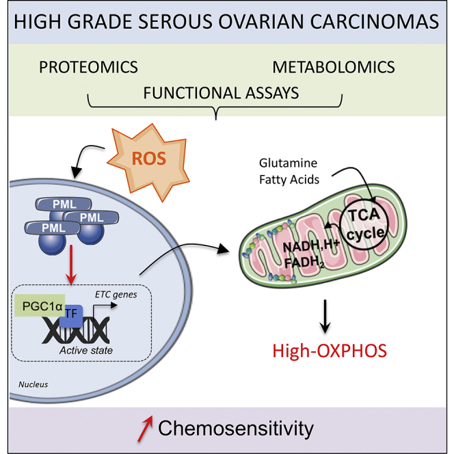
          High-grade serous ovarian cancer (HGSOC) remains an unmet medical challenge. Here, we unravel an unanticipated metabolic heterogeneity in HGSOC. By combining proteomic, metabolomic, and bioergenetic analyses, we identify two molecular subgroups, low- and high-OXPHOS. While low-OXPHOS exhibit a glycolytic metabolism, high-OXPHOS HGSOCs rely on oxidative phosphorylation, supported by glutamine and fatty acid oxidation, and show chronic oxidative stress. We identify an important role for the PML-PGC-1α axis in the metabolic features of high-OXPHOS HGSOC. In high-OXPHOS tumors, chronic oxidative stress promotes aggregation of PML-nuclear bodies, resulting in activation of the transcriptional co-activator PGC-1α. Active PGC-1α increases synthesis of electron transport chain complexes, thereby promoting mitochondrial respiration. Importantly, high-OXPHOS HGSOCs exhibit increased response to conventional chemotherapies, in which increased oxidative stress, PML, and potentially ferroptosis play key functions. Collectively, our data establish a stress-mediated PML-PGC-1α-dependent mechanism that promotes OXPHOS metabolism and chemosensitivity in ovarian cancer.

          Graphical Abstract

          Highlights

          • High-grade serous ovarian cancers display OXPHOS metabolic heterogeneity

          • High-OXPHOS show high electron transport chain synthesis and respiration rate

          • Oxidative stress in high-OXPHOS HGSOC activates PML-PGC-1α and ETC transcription

          • High-OXPHOS HGSOCs show enhanced chemosensitivity through oxidative stress and PML

          Abstract

          Gentric et al. identify metabolically heterogeneous OXPHOS subgroups in high-grade serous ovarian cancers (HGSOCs). In high-OXPHOS tumors, chronic oxidative stress promotes aggregation of PML-nuclear bodies that activate PGC-1α, electron transport chain synthesis, and mitochondrial respiration. High-OXPHOS HGSOCs exhibit chemosensitivity, in which increased oxidative stress and PML play key functions.

          Related collections

          Most cited references36

          • Record: found
          • Abstract: found
          • Article: found

          MYC/PGC-1α Balance Determines the Metabolic Phenotype and Plasticity of Pancreatic Cancer Stem Cells.

          The anti-diabetic drug metformin targets pancreatic cancer stem cells (CSCs), but not their differentiated progenies (non-CSCs), which may be related to distinct metabolic phenotypes. Here we conclusively demonstrate that while non-CSCs were highly glycolytic, CSCs were dependent on oxidative metabolism (OXPHOS) with very limited metabolic plasticity. Thus, mitochondrial inhibition, e.g., by metformin, translated into energy crisis and apoptosis. However, resistant CSC clones eventually emerged during treatment with metformin due to their intermediate glycolytic/respiratory phenotype. Mechanistically, suppression of MYC and subsequent increase of PGC-1α were identified as key determinants for the OXPHOS dependency of CSCs, which was abolished in resistant CSC clones. Intriguingly, no resistance was observed for the mitochondrial ROS inducer menadione and resistance could also be prevented/reversed for metformin by genetic/pharmacological inhibition of MYC. Thus, the specific metabolic features of pancreatic CSCs are amendable to therapeutic intervention and could provide the basis for developing more effective therapies to combat this lethal cancer.
            Bookmark
            • Record: found
            • Abstract: found
            • Article: found
            Is Open Access

            Patterns of genomic loss of heterozygosity predict homologous recombination repair defects in epithelial ovarian cancer

            Background: Defects in BRCA1, BRCA2, and other members of the homologous recombination pathway have potential therapeutic relevance when used to support agents that introduce or exploit double-stranded DNA breaks. This study examines the association between homologous recombination defects and genomic patterns of loss of heterozygosity (LOH). Methods: Ovarian tumours from two independent data sets were characterised for defects in BRCA1, BRCA2, and RAD51C, and LOH profiles were generated. Publically available data were downloaded for a third independent data set. The same analyses were performed on 57 cancer cell lines. Results: Loss of heterozygosity regions of intermediate size were observed more frequently in tumours with defective BRCA1 or BRCA2 (P=10−11). The homologous recombination deficiency (HRD) score was defined as the number of these regions observed in a tumour sample. The association between HRD score and BRCA deficiency was validated in two independent ovarian cancer data sets (P=10−5 and 10−29), and identified breast and pancreatic cell lines with BRCA defects. Conclusion: The HRD score appears capable of detecting homologous recombination defects regardless of aetiology or mechanism. This score could facilitate the use of PARP inhibitors and platinum in breast, ovarian, and other cancers.
              Bookmark
              • Record: found
              • Abstract: found
              • Article: not found

              PPARγ agonists induce a white-to-brown fat conversion through stabilization of PRDM16 protein.

              Brown adipose tissue dissipates energy through heat and functions as a defense against cold and obesity. PPARγ ligands have been shown to induce the browning of white adipocytes; however, the underlying mechanisms remain unclear. Here, we show that PPARγ ligands require full agonism to induce a brown fat gene program preferentially in subcutaneous white adipose. These effects require expression of PRDM16, a factor that controls the development of classical brown fat. Depletion of PRDM16 blunts the effects of the PPARγ agonist rosiglitazone on the induced brown fat gene program. Conversely, PRDM16 and rosiglitazone synergistically activate the brown fat gene program in vivo. This synergy is tightly associated with an increased accumulation of PRDM16 protein, due in large measure to an increase in the half-life of the protein in agonist treated cells. Identifying compounds that stabilize PRDM16 protein may represent a plausible therapeutic pathway for the treatment of obesity and diabetes. Copyright © 2012 Elsevier Inc. All rights reserved.
                Bookmark

                Author and article information

                Contributors
                Journal
                Cell Metab
                Cell Metab
                Cell Metabolism
                Cell Press
                1550-4131
                1932-7420
                08 January 2019
                08 January 2019
                : 29
                : 1
                : 156-173.e10
                Affiliations
                [1 ]Institut Curie, Stress and Cancer Laboratory, Equipe Labelisée par la Ligue Nationale contre le Cancer, PSL Research University, 26, rue d’Ulm, 75005 Paris, France
                [2 ]Inserm, U830, 26, rue d’Ulm, Paris 75005, France
                [3 ]Translational Research Department, Laboratory of Preclinical Investigation, Institut Curie, 26, rue d’Ulm, Paris 75248, France
                [4 ]Institut Curie, PSL Research University, CNRS, UMR 144, 75005 Paris, France
                [5 ]Structure and Membrane Compartments, CNRS, UMR 144, 75005 Paris, France
                [6 ]Cell and Tissue Imaging Core Facility PICT-IBiSA, Institut Curie, 75248 Paris, France
                [7 ]DNA Repair and Uveal Melanoma (D.R.U.M.) team, Institut Curie, 26, rue d’Ulm, 75248 Paris Cedex 05, France
                [8 ]Collège de France, PSL Research University, Place Marcellin Berthelot, 75005 Paris, France
                [9 ]Inserm UMR 944, Equipe Labellisée par la Ligue Nationale contre le Cancer, Paris Diderot University, Hôpital St. Louis, Paris, France
                [10 ]Chemical Biology of Cancer, CNRS UMR3666, Inserm U1143, Institut Curie, Equipe Labellisée par la Ligue Nationale contre le Cancer, PSL Research University, 26, rue d’Ulm, Paris 75248, France
                [11 ]Department of Pathology, Institut Curie Hospital, 26, rue d’Ulm, 75248 Paris, France
                [12 ]Inserm U1211, Université de Bordeaux, 146 rue Léo Saignat, 33000 Bordeaux, France
                Author notes
                []Corresponding author geraldine.gentric@ 123456curie.fr
                [∗∗ ]Corresponding author fatima.mechta-grigoriou@ 123456curie.fr
                [13]

                Lead Contact

                Article
                S1550-4131(18)30567-9
                10.1016/j.cmet.2018.09.002
                6331342
                30244973
                f684cc4b-4189-48c3-98aa-ae0967d8f173
                © 2018 The Authors

                This is an open access article under the CC BY license (http://creativecommons.org/licenses/by/4.0/).

                History
                : 11 January 2018
                : 11 July 2018
                : 31 August 2018
                Categories
                Article

                Cell biology
                oxphos,reactive oxygen species,promyelocytic leukemia protein,peroxisome proliferator-activated receptor gamma coactivator-1α,pgc-1α,response to treatment,ovarian cancer,oxidative stress,chemoresistance,ferroptosis

                Comments

                Comment on this article