1
views
0
recommends
+1 Recommend
0 collections
    0
    shares
      • Record: found
      • Abstract: found
      • Article: found
      Is Open Access

      Ruptured Median Raphe Cyst Mimicking a Vascular Penile Mass on Ultrasound

      case-report
      1 , , 1 , 1 , 2
      Case Reports in Radiology
      Hindawi

      Read this article at
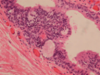
      Bookmark
          There is no author summary for this article yet. Authors can add summaries to their articles on ScienceOpen to make them more accessible to a non-specialist audience.

          Abstract

          Median raphe cysts are uncommon benign cysts thought to occur due to improper fusion of the genital tubercle and can occur anywhere along the median raphe, from the glans to the anus, most commonly occurring along the ventral penile shaft. Limited information is available in the literature about the common imaging features of median raphe cysts with available reports highlighting an avascular cystic lesion. Our case demonstrates a 10-year-old male patient presenting with a ventral penile mass that demonstrated interval growth in the absence of trauma without overlying skin changes. Doppler ultrasound examination demonstrated a solid vascular mass measuring up to 1.6 cm at the ventral aspect of the penis with arterial and venous waveforms. The patient underwent elective resection of the mass which revealed a 2.0 cm inflamed glandular subtype median raphe cyst. This report demonstrates an atypical imaging presentation of an inflamed median raphe cyst, particularly that of a heterogeneous solid mass with arterial and venous blood flow on ultrasound.

          Related collections

          Most cited references9

          • Record: found
          • Abstract: found
          • Article: found
          Is Open Access

          Male median raphe cysts: serial retrospective analysis and histopathological classification

          Background To review the clinical and pathological characteristics of median raphe cysts and to classify the lesions according to pathogenesis and histopathological findings. Methods The medical records of patients who were diagnosed with median raphe cysts between 2001 and 2010 were reviewed to document the clinical presentation and pathological findings of the cysts. Results Most patients were asymptomatic; however, 9 patients had inflammatory or infectious cysts that were tender or painful. Four patients who had cysts on the parameatus and distal prepuce had difficulty voiding. Hematuria and hematospermia were noted in 2 cases. Thirty-one cysts were lined with an urothelium-like epithelium, and a squamous epithelium lining was found in 3 cases. In 2 cases, a well-formed mucinous glandular structure was observed. The other 20 cysts consisted of mixed epithelia. After excision of the cysts under local or general anesthesia, an urethral fistula developed as a complication in only 1 case. Conclusions Median raphe cysts are benign lesions formed due to tissue trapping during the development of urethral folds. The cysts can be defined into 4 types based on pathological findings: urethral, epidermoid, glandular, and mixed. The associated symptoms and signs should be taken into consideration when determining the treatment for the cysts. Virtual slides The virtual slide(s) for this article can be found here: http//http://www.diagnosticpathology.diagnomx.eu/vs/7727074877500751
            Bookmark
            • Record: found
            • Abstract: found
            • Article: found
            Is Open Access

            Median raphe cyst of the penis: a case report and review of the literature

            Background A defect in embryological development or closure of median raphe may lead to formation of cyst(s) anywhere in the midline from glans to anus. These cysts are referred to as median raphe cysts, an uncommonly encountered clinical condition. The cyst is generally solitary, with the penile shaft being the most common location, with average size of around 1 cm. The diagnosis is mostly clinical and confirmed histologically. We report a case of a patient with a rare histological variant of median raphe cyst and provide a focused review on presentation, histopathology, and management. Case presentation A 29-year-old unmarried Nepali man presented to our clinic with an asymptomatic, solitary, soft, translucent, nontender cystic lesion of about 1-cm diameter at the ventral aspect of glans penis, close to the meatus, that had been noticed at the age of 3 and was nonprogressive for the past 15 years. Ultrasonography demonstrated an isoechoic cystic lesion at the tip of the penis, separated from the urethra, and lying entirely within the mucosa without any evidence of solid component, septation, or vascularity. On the basis of clinical and ultrasonographic findings, a diagnosis of median raphe cyst of the penis was made. The cyst was excised with the patient under local anesthesia, and there was no evidence of recurrence in 2 years of follow-up. The histopathological examination with Hematoxylin and eosin staining showed the cyst wall was lined partly by ciliated pseudostratified columnar epithelium and partly by columnar epithelium with apical mucin. Conclusions Median raphe cyst is an uncommon, mostly asymptomatic condition in young patients. The cyst may occur anywhere along the midline from glans to anus. The diagnosis is clinical with histological confirmation. Excision is the treatment of choice with minimal chance of recurrence.
              Bookmark
              • Record: found
              • Abstract: found
              • Article: not found

              A case of epidermoid median raphe cyst traversing the corpora cavernosa.

              Median raphe cysts are congenital lesions that typically have a superficial appearance. We present a very unusual case of a deep perineal mass in a six-year-old boy. The lesion extends into the corpus cavernosum, suggesting that the anomaly was an early embryological event. Histopathological features are consistent with an epidermoid type of median raphe cyst.
                Bookmark

                Author and article information

                Contributors
                Journal
                Case Rep Radiol
                Case Rep Radiol
                CRIRA
                Case Reports in Radiology
                Hindawi
                2090-6862
                2090-6870
                2022
                28 February 2022
                : 2022
                : 8899541
                Affiliations
                1Department of Diagnostic Radiology, Beaumont Hospital, Royal Oak, MI, Michigan, USA
                2Michigan State University College of Osteopathic Medicine, East Lansing, MI, Michigan, USA
                Author notes

                Academic Editor: Daniel P. Link

                Author information
                https://orcid.org/0000-0001-7983-3192
                Article
                10.1155/2022/8899541
                8901341
                f5f5654a-ee9c-4204-a14c-60e15a59a605
                Copyright © 2022 Amir Pakray et al.

                This is an open access article distributed under the Creative Commons Attribution License, which permits unrestricted use, distribution, and reproduction in any medium, provided the original work is properly cited.

                History
                : 19 September 2020
                : 7 December 2021
                : 7 February 2022
                Categories
                Case Report

                Radiology & Imaging
                Radiology & Imaging

                Comments

                Comment on this article