11
views
0
recommends
+1 Recommend
0 collections
    0
    shares
      • Record: found
      • Abstract: found
      • Article: found
      Is Open Access

      Evaluation of retrobulbar circulation in type 2 diabetic patients using color Doppler imaging

      research-article

      Read this article at

      Bookmark
          There is no author summary for this article yet. Authors can add summaries to their articles on ScienceOpen to make them more accessible to a non-specialist audience.

          Abstract

          Purpose:

          To investigate the retrobulbar circulatory parameters in type 2 diabetes mellitus patients with color Doppler imaging (CDI) and compare the results with nondiabetic controls.

          Methods:

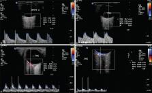
          This prospective study included 50 type 2 diabetic patients and 50 age-matched controls. Seven field stereo fundus photography was used to diagnose and classify diabetic retinopathy (DR). Diabetic patients were further divided into two: Group 1, consisted of patients with no DR, mild and moderate non-proliferative DR ( n = 36); Group 2, severe nonproliferative and proliferative DR ( n = 14). CDI was performed using Philips iU22 xMATRIX ultrasound. The peak systolic velocity (PSV), end-diastolic velocity (EDV), resistivity index (RI) and pulsatile index (PI) of ophthalmic (OA), posterior ciliary artery (PCA), and central retinal artery (CRA) along with central retinal vein (CRV) were recorded.

          Results:

          RI in the ophthalmic artery was significantly higher in both DR groups than the control group ( P = 0.000). Diabetic Group 1 had decreased blood flow velocity (PSV and EDV) in PCA compared to controls ( P = 0.046 and P = 0.010, respectively). Group 2 DR had significantly reduced EDV and increased RI in CRA compared to Group 1 ( P = 0.015). Binary logistic regression analysis revealed glycosylated hemoglobin and RI of OA to be independent risk factors of DR.

          Conclusion:

          Significant changes in resistivity index and flow velocities were observed in the retrobulbar vessels, especially in ophthalmic artery in diabetics compared to controls. CDI with results of increased resistance or decreased flow could be useful to predict individuals at higher risk for developing severe DR.

          Related collections

          Most cited references26

          • Record: found
          • Abstract: found
          • Article: not found

          Vascular endothelial growth factors and angiogenesis in eye disease.

          The vascular endothelial growth factor (VEGF) family of growth factors controls pathological angiogenesis and increased vascular permeability in important eye diseases such as diabetic retinopathy (DR) and age-related macular degeneration (AMD). The purpose of this review is to develop new insights into the cell biology of VEGFs and vascular cells in angiogenesis and vascular leakage in general, and to provide the rationale and possible pitfalls of inhibition of VEGFs as a therapy for ocular disease. From the literature it is clear that overexpression of VEGFs and their receptors VEGFR-1, VEGFR-2 and VEGFR-3 is causing increased microvascular permeability and angiogenesis in eye conditions such as DR and AMD. When we focus on the VEGF receptors, recent findings suggest a role of VEGFR-1 as a functional receptor for placenta growth factor (PlGF) and vascular endothelial growth factor-A (VEGF)-A in pericytes and vascular smooth muscle cells in vivo rather than in endothelial cells, and strongly suggest involvement of pericytes in early phases of angiogenesis. In addition, the evidence pointing to distinct functions of VEGFs in physiology in and outside the vasculature is reviewed. The cellular distribution of VEGFR-1, VEGFR-2 and VEGFR-3 suggests various specific functions of the VEGF family in normal retina, both in the retinal vasculature and in neuronal elements. Furthermore, we focus on recent findings that VEGFs secreted by epithelia, including the retinal pigment epithelium (RPE), are likely to mediate paracrine vascular survival signals for adjacent endothelia. In the choroid, derailment of this paracrine relation and overexpression of VEGF-A by RPE may explain the pathogenesis of subretinal neovascularisation in AMD. On the other hand, this paracrine relation and other physiological functions of VEGFs may be endangered by therapeutic VEGF inhibition, as is currently used in several clinical trials in DR and AMD.
            Bookmark
            • Record: found
            • Abstract: found
            • Article: not found

            Investigating the choriocapillaris and choroidal vasculature with new optical coherence tomography technologies.

            The body of knowledge of in vivo investigation of the choroid has been markedly enhanced by recent technological advances in optical coherence tomography (OCT). New insights elucidating the morphological features of the choriocapillaris and choroidal vasculature, in both physiological and pathological conditions, indicate that the choroid plays a pivotal role in many posterior segment diseases. In this article, a review of the histological characteristics of the choroid, which must be considered for the proper interpretation of in vivo imaging, is followed by a comprehensive discussion of fundamental principles of the current state-of-the-art in OCT, including cross-sectional OCT, en face OCT, and OCT angiography using both spectral domain OCT and swept source OCT technologies. A detailed review of the tomographic features of the choroid in the normal eye is followed by relevant findings in prevalent chorioretinal diseases, focusing on major causes of vision loss such as typical early and advanced age-related macular degeneration, polypoidal choroidal vasculopathy, central serous chorioretinopathy, pachychoroid spectrum disorders, diabetic choroidopathy, and myopia.
              Bookmark
              • Record: found
              • Abstract: found
              • Article: not found

              Microvascular lesions of diabetic retinopathy: clues towards understanding pathogenesis?

              Retinopathy is a major complication of diabetes mellitus and this condition remains a leading cause of blindness in the working population of developed countries. As diabetic retinopathy progresses a range of neuroglial and microvascular abnormalities develop although it remains unclear how these pathologies relate to each other and their net contribution to retinal damage. From a haemodynamic perspective, evidence suggests that there is an early reduction in retinal perfusion before the onset of diabetic retinopathy followed by a gradual increase in blood flow as the complication progresses. The functional reduction in retinal blood flow observed during early diabetic retinopathy may be additive or synergistic to pro-inflammatory changes, leucostasis and vaso-occlusion and thus be intimately linked to the progressive ischaemic hypoxia and increased blood flow associated with later stages of the disease. In the current review a unifying framework is presented that explains how arteriolar dysfunction and haemodynamic changes may contribute to late stage microvascular pathology and vision loss in human diabetic retinopathy.
                Bookmark

                Author and article information

                Journal
                Indian J Ophthalmol
                Indian J Ophthalmol
                IJO
                Indian Journal of Ophthalmology
                Wolters Kluwer - Medknow (India )
                0301-4738
                1998-3689
                June 2020
                25 May 2020
                : 68
                : 6
                : 1108-1114
                Affiliations
                [1]Department of Ophthalmology, PSG Institute of Medical Sciences and Research, Coimbatore, Tamil Nadu, India
                [1 ]Department of Radiology, PSG Institute of Medical Sciences and Research, Coimbatore, Tamil Nadu, India
                Author notes
                Correspondence to: Dr. Vikrant Kanagaraju, Department of Radiology, PSG Institute of Medical Sciences and Research, Coimbatore - 641 004, Tamil Nadu, India. E-mail: vikrantmailin@ 123456gmail.com
                Article
                IJO-68-1108
                10.4103/ijo.IJO_1398_19
                7508083
                32461442
                f4fc467c-9c76-4188-9fa0-7fb149c7ce8e
                Copyright: © 2020 Indian Journal of Ophthalmology

                This is an open access journal, and articles are distributed under the terms of the Creative Commons Attribution-NonCommercial-ShareAlike 4.0 License, which allows others to remix, tweak, and build upon the work non-commercially, as long as appropriate credit is given and the new creations are licensed under the identical terms.

                History
                : 30 July 2019
                : 01 November 2019
                : 28 December 2019
                Categories
                Original Article

                Ophthalmology & Optometry
                blood flow velocities,color doppler ultrasonography,diabetes mellitus,retrobulbar

                Comments

                Comment on this article