103
views
0
recommends
+1 Recommend
0 collections
    0
    shares
      • Record: found
      • Abstract: found
      • Article: found
      Is Open Access

      Misnomers in Dermatology: An Update

      other

      Read this article at

      Bookmark
          There is no author summary for this article yet. Authors can add summaries to their articles on ScienceOpen to make them more accessible to a non-specialist audience.

          Abstract

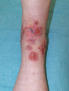
          The name of a condition in dermatology, gives a clue regarding the clinical feature, etiology, or histopathology of the disease. A disease might have been termed wrongly due to its resemblance to another known condition. Misnomers often mislead a physician regarding the etiology or histopathology of the condition. Here is a list of misnomers, with explanation, and the appropriate name in parentheses.

          Related collections

          Most cited references73

          • Record: found
          • Abstract: found
          • Article: not found

          Hemangiomas and vascular malformations in infants and children: a classification based on endothelial characteristics.

          Forty-nine specimens from a variety of vascular lesions were analyzed for cellular characteristics. Two major categories of lesions emerged from this investigation: hemangiomas and vascular malformations. This classification and its implications are justified by several considerations. Hemangiomas in the proliferating phase (n = 14) were distinguished by (1) endothelial hyperplasia with incorporation of [3H]thymidine, (2) multilaminated basement membrane formation beneath the endothelium, and (3) clinical history of rapid growth during early infancy. Hemangiomas in the involuting phase (n = 12) exhibited (1) histologic fibrosis and fat deposition, (2) low to absent [3H]thymidine labeling of endothelial cells, and (3) rapid growth and subsequent regression. The endothelium in hemangiomas had many characteristics of differentiation: Weibel-Palade bodies, alkaline phosphatase, and factor VIII production. Vascular malformations (n = 23) demonstrated no tritiated thymidine incorporation and normal ultrastructural characteristics. These lesions were usually noted at birth, grew proportionately with the child, and consisted of abnormal, often combined, capillary, arterial, venous, and lymphatic vascular elements. This cell-oriented analysis provides a simple yet comprehensive classification of vascular lesions of infancy and childhood and serves as a guide for diagnosis, management, and further research.
            Bookmark
            • Record: found
            • Abstract: found
            • Article: not found

            Cellular markers that distinguish the phases of hemangioma during infancy and childhood.

            Hemangiomas, localized tumors of blood vessels, appear in approximately 10-12% of Caucasian infants. These lesions are characterized by a rapid proliferation of capillaries for the first year (proliferating phase), followed by slow, inevitable, regression of the tumor over the ensuing 1-5 yr (involuting phase), and continual improvement until 6-12 yr of age (involuted phase). To delineate the clinically observed growth phases of hemangiomas at a cellular level, we undertook an immunohistochemical analysis using nine independent markers. The proliferating phase was defined by high expression of proliferating cell nuclear antigen, type IV collagenase, and vascular endothelial growth factor. Elevated expression of the tissue inhibitor of metalloproteinase, TIMP 1, an inhibitor of new blood vessel formation, was observed exclusively in the involuting phase. High expression of basic fibroblast growth factor (bFGF) and urokinase was present in the proliferating and involuting phases. There was coexpression of bFGF and endothelial phenotypic markers CD31 and von Willebrand factor in the proliferating phase. These results provide an objective basis for staging hemangiomas and may be used to evaluate pharmacological agents, such as corticosteroids and interferon alfa-2a, which accelerate regression of hemangiomas. By contrast, vascular malformations do not express proliferating cell nuclear antigen, vascular endothelial growth factor, bFGF, type IV collagenase, and urokinase. These data demonstrate immunohistochemical differences between proliferating hemangiomas and vascular malformations which reflect the biological distinctions between these vascular lesions.
              Bookmark
              • Record: found
              • Abstract: found
              • Article: found
              Is Open Access

              Pyoderma gangrenosum – a review

              Pyoderma gangrenosum (PG) is a rare noninfectious neutrophilic dermatosis. Clinically it starts with sterile pustules that rapidly progress and turn into painful ulcers of variable depth and size with undermined violaceous borders. The legs are most commonly affected but other parts of the skin and mucous membranes may also be involved. Course can be mild or malignant, chronic or relapsing with remarkable morbidity. In many cases PG is associated with an underlying disease, most commonly inflammatory bowel disease, rheumatic or haematological disease and malignancy. Diagnosis of PG is based on history of an underlying disease, typical clinical presentation, histopathology, and exclusion of other diseases that would lead to a similar appearance. The peak of incidence occurs between the ages of 20 to 50 years with women being more often affected than men. Aetiology has not been clearly determined yet. The treatment of PG is a challenge. Randomized, double-blinded prospective multicenter trials for PG are not available. The best documented treatments are systemic corticosteroids and ciclosporin A. Combinations of steroids with cytotoxic drugs are used in resistant cases. The combination of steroids with sulfa drugs or immunosuppressants has been used as steroid-sparing modalities. Anti-tumor necrosis alpha therapy in Crohn's disease showed a rapid response of PG. Skin transplants and the application of bioengineered skin is useful in selected cases as a complement to the immunosuppressive treatment. Topical therapy with modern wound dressings is useful to minimize pain and the risk of secondary infections. Despite recent advances in therapy, the prognosis of PG remains unpredictable.
                Bookmark

                Author and article information

                Journal
                Indian J Dermatol
                Indian J Dermatol
                IJD
                Indian Journal of Dermatology
                Medknow Publications & Media Pvt Ltd (India )
                0019-5154
                1998-3611
                Nov-Dec 2013
                : 58
                : 6
                : 467-474
                Affiliations
                [1] From the Department of Dermatology, STD, Leprosy, Bangalore Medical College and Research Institute, Bangalore, India
                Author notes
                Address for correspondence: Dr. Somaiah A Savitha, No 52, OPD B Block, Department of Dermatology, STD and Leprosy, Bangalore Medical College and Research Institute, Bangalore, India. drsavithasomaiah@ 123456gmail.com
                Article
                IJD-58-467
                10.4103/0019-5154.119962
                3827521
                24249901
                f4d5cce2-3e86-4d49-b2b6-92dceccb3f73
                Copyright: © Indian Journal of Dermatology

                This is an open-access article distributed under the terms of the Creative Commons Attribution-Noncommercial-Share Alike 3.0 Unported, which permits unrestricted use, distribution, and reproduction in any medium, provided the original work is properly cited.

                History
                : September 2012
                : December 2012
                Categories
                Resident's Page

                Dermatology
                dermatology,misnomers,synonyms
                Dermatology
                dermatology, misnomers, synonyms

                Comments

                Comment on this article