64
views
0
recommends
+1 Recommend
0 collections
    0
    shares
      • Record: found
      • Abstract: found
      • Article: found
      Is Open Access

      Oral N-acetylcysteine decreases IFN-γ production and ameliorates ischemia-reperfusion injury in steatotic livers

      research-article

      Read this article at

      Bookmark
          There is no author summary for this article yet. Authors can add summaries to their articles on ScienceOpen to make them more accessible to a non-specialist audience.

          Abstract

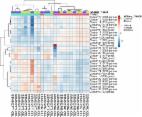
          Type 1 Natural Killer T-cells (NKT1 cells) play a critical role in mediating hepatic ischemia-reperfusion injury (IRI). Although hepatic steatosis is a major risk factor for preservation type injury, how NKT cells impact this is understudied. Given NKT1 cell activation by phospholipid ligands recognized presented by CD1d, we hypothesized that NKT1 cells are key modulators of hepatic IRI because of the increased frequency of activating ligands in the setting of hepatic steatosis. We first demonstrate that IRI is exacerbated by a high-fat diet (HFD) in experimental murine models of warm partial ischemia. This is evident in the evaluation of ALT levels and Phasor-Fluorescence Lifetime (Phasor-FLIM) Imaging for glycolytic stress. Polychromatic flow cytometry identified pronounced increases in CD45+CD3+NK1.1+NKT1 cells in HFD fed mice when compared to mice fed a normal diet (ND). This observation is further extended to IRI, measuring ex vivo cytokine expression in the HFD and ND. Much higher interferon-gamma (IFN-γ) expression is noted in the HFD mice after IRI. We further tested our hypothesis by performing a lipidomic analysis of hepatic tissue and compared this to Phasor-FLIM imaging using “long lifetime species”, a byproduct of lipid oxidation. There are higher levels of triacylglycerols and phospholipids in HFD mice. Since N-acetylcysteine (NAC) is able to limit hepatic steatosis, we tested how oral NAC supplementation in HFD mice impacted IRI. Interestingly, oral NAC supplementation in HFD mice results in improved hepatic enhancement using contrast-enhanced magnetic resonance imaging (MRI) compared to HFD control mice and normalization of glycolysis demonstrated by Phasor-FLIM imaging. This correlated with improved biochemical serum levels and a decrease in IFN-γ expression at a tissue level and from CD45+CD3+CD1d+ cells. Lipidomic evaluation of tissue in the HFD+NAC mice demonstrated a drastic decrease in triacylglycerol, suggesting downregulation of the PPAR-γ pathway.

          Related collections

          Most cited references70

          • Record: found
          • Abstract: found
          • Article: found
          Is Open Access

          ClustVis: a web tool for visualizing clustering of multivariate data using Principal Component Analysis and heatmap

          The Principal Component Analysis (PCA) is a widely used method of reducing the dimensionality of high-dimensional data, often followed by visualizing two of the components on the scatterplot. Although widely used, the method is lacking an easy-to-use web interface that scientists with little programming skills could use to make plots of their own data. The same applies to creating heatmaps: it is possible to add conditional formatting for Excel cells to show colored heatmaps, but for more advanced features such as clustering and experimental annotations, more sophisticated analysis tools have to be used. We present a web tool called ClustVis that aims to have an intuitive user interface. Users can upload data from a simple delimited text file that can be created in a spreadsheet program. It is possible to modify data processing methods and the final appearance of the PCA and heatmap plots by using drop-down menus, text boxes, sliders etc. Appropriate defaults are given to reduce the time needed by the user to specify input parameters. As an output, users can download PCA plot and heatmap in one of the preferred file formats. This web server is freely available at http://biit.cs.ut.ee/clustvis/.
            Bookmark
            • Record: found
            • Abstract: found
            • Article: found
            Is Open Access

            MetaboAnalyst 4.0: towards more transparent and integrative metabolomics analysis

            Abstract We present a new update to MetaboAnalyst (version 4.0) for comprehensive metabolomic data analysis, interpretation, and integration with other omics data. Since the last major update in 2015, MetaboAnalyst has continued to evolve based on user feedback and technological advancements in the field. For this year's update, four new key features have been added to MetaboAnalyst 4.0, including: (1) real-time R command tracking and display coupled with the release of a companion MetaboAnalystR package; (2) a MS Peaks to Pathways module for prediction of pathway activity from untargeted mass spectral data using the mummichog algorithm; (3) a Biomarker Meta-analysis module for robust biomarker identification through the combination of multiple metabolomic datasets and (4) a Network Explorer module for integrative analysis of metabolomics, metagenomics, and/or transcriptomics data. The user interface of MetaboAnalyst 4.0 has been reengineered to provide a more modern look and feel, as well as to give more space and flexibility to introduce new functions. The underlying knowledgebases (compound libraries, metabolite sets, and metabolic pathways) have also been updated based on the latest data from the Human Metabolome Database (HMDB). A Docker image of MetaboAnalyst is also available to facilitate download and local installation of MetaboAnalyst. MetaboAnalyst 4.0 is freely available at http://metaboanalyst.ca.
              Bookmark
              • Record: found
              • Abstract: found
              • Article: not found

              The phasor approach to fluorescence lifetime imaging analysis.

              Changing the data representation from the classical time delay histogram to the phasor representation provides a global view of the fluorescence decay at each pixel of an image. In the phasor representation we can easily recognize the presence of different molecular species in a pixel or the occurrence of fluorescence resonance energy transfer. The analysis of the fluorescence lifetime imaging microscopy (FLIM) data in the phasor space is done observing clustering of pixels values in specific regions of the phasor plot rather than by fitting the fluorescence decay using exponentials. The analysis is instantaneous since is not based on calculations or nonlinear fitting. The phasor approach has the potential to simplify the way data are analyzed in FLIM, paving the way for the analysis of large data sets and, in general, making the FLIM technique accessible to the nonexpert in spectroscopy and data analysis.
                Bookmark

                Author and article information

                Contributors
                Journal
                Front Immunol
                Front Immunol
                Front. Immunol.
                Frontiers in Immunology
                Frontiers Media S.A.
                1664-3224
                05 September 2022
                2022
                : 13
                : 898799
                Affiliations
                [1] 1 MedStar Georgetown Transplant Institute, MedStar Georgetown University Hospital and the Center for Translational Transplant Medicine, Georgetown University Medical Center , Washington, DC, United States
                [2] 2 Department of Surgery, Naval Medical Center Portsmouth , Portsmouth, VA, United States
                [3] 3 Department of Biochemistry and Molecular & Cellular Biology, Georgetown University , Washington, DC, United States
                [4] 4 Microscopy & Imaging Shared Resource, Georgetown University , Washington, DC, United States
                [5] 5 Center for Translational Imaging, Georgetown University Medical Center , Washington, DC, United States
                [6] 6 Department of Oncology, Lombardi Comprehensive Cancer Center, Georgetown University Medical Center , Washington, DC, United States
                [7] 7 Department of Pathology, MedStar Georgetown University Hospital , Washington, DC, United States
                [8] 8 Division of Endocrinology, Metabolism, & Diabetes, University of Colorado Anschutz Medical Campus , Aurora, CO, United States
                [9] 9 Departments of Anesthesiology and Medicine, Beth Israel Deaconess Medical Center, Harvard Medical School , Boston, MA, United States
                [10] 10 Department of Radiology, MedStar Georgetown University Hospital , Washington, DC, United States
                Author notes

                Edited by: Tao Qiu, Renmin Hospital of Wuhan University, China

                Reviewed by: JingHong Wan, INSERM U1149 Centre de Recherche sur l’Inflammation, France; Lan Wu, Vanderbilt University Medical Center, United States

                †These authors have contributed equally to this work and share first authorship

                This article was submitted to Molecular Innate Immunity, a section of the journal Frontiers in Immunology

                Article
                10.3389/fimmu.2022.898799
                9486542
                36148239
                f4a76104-62ef-45cf-8692-1d5fd1281caf
                Copyright © 2022 Liggett, Kang, Ranjit, Rodriguez, Loh, Patil, Cui, Duttargi, Nguyen, He, Lee, Oza, Frank, Kwon, Li, Kallakury, Libby, Levi, Robson, Fishbein, Cui, Albanese, Khan and Kroemer

                This is an open-access article distributed under the terms of the Creative Commons Attribution License (CC BY). The use, distribution or reproduction in other forums is permitted, provided the original author(s) and the copyright owner(s) are credited and that the original publication in this journal is cited, in accordance with accepted academic practice. No use, distribution or reproduction is permitted which does not comply with these terms.

                History
                : 17 March 2022
                : 11 July 2022
                Page count
                Figures: 8, Tables: 0, Equations: 2, References: 71, Pages: 20, Words: 9888
                Funding
                Funded by: National Institutes of Health , doi 10.13039/100000002;
                Funded by: National Institutes of Health , doi 10.13039/100000002;
                Funded by: National Institutes of Health , doi 10.13039/100000002;
                Funded by: National Institutes of Health , doi 10.13039/100000002;
                Funded by: National Institutes of Health , doi 10.13039/100000002;
                Funded by: National Institutes of Health , doi 10.13039/100000002;
                Categories
                Immunology
                Original Research

                Immunology
                nkt (natural killer t) cell,hepatic steatosis,ifn-gamma,ischemia-reperfusion injury (iri),n-acetylcysteine (nac),phasor-flim,gadoxetate disodium,liver transplantation

                Comments

                Comment on this article

                scite_
                0
                0
                0
                0
                Smart Citations
                0
                0
                0
                0
                Citing PublicationsSupportingMentioningContrasting
                View Citations

                See how this article has been cited at scite.ai

                scite shows how a scientific paper has been cited by providing the context of the citation, a classification describing whether it supports, mentions, or contrasts the cited claim, and a label indicating in which section the citation was made.

                Similar content36

                Cited by9

                Most referenced authors1,019