18
views
0
recommends
+1 Recommend
0 collections
    0
    shares
      • Record: found
      • Abstract: found
      • Article: found
      Is Open Access

      Surface plasmon resonance immunosensor for label-free detection of BIRC5 biomarker in spontaneously occurring canine mammary tumours

      research-article

      Read this article at

      Bookmark
          There is no author summary for this article yet. Authors can add summaries to their articles on ScienceOpen to make them more accessible to a non-specialist audience.

          Abstract

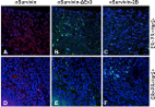
          We report detection of Baculoviral inhibitor of apoptosis repeat containing-5 (BIRC5) protein biomarker in dog serum by label-free surface plasmon resonance (SPR) immunosensor. Initially, overexpression of BIRC5 in canine mammary tumour (CMT) tissues was confirmed by real-time PCR. Recombinant BIRC5 was produced and protein specific antibodies developed in guinea pig specifically reacted with native protein in immunohistochemistry and immunocytochemistry. SPR immunosensor was developed by fabricating anti-BIRC5 antibodies on gold sensor disc. The equilibrium dissociation constant, (K D = k d/k a) was 12.1 × 10 −12 M; which indicates that antibodies are of high affinity with sensitivity in picomolar range. The SPR assay could detect as low as 6.25 pg/ml of BIRC5 protein in a calibration experiment (r 2 = 0.9964). On testing real clinical samples, 95% specificity and 73.33% sensitivity were recorded. The average amount of serum BIRC5 in dogs with CMT was 110.02 ± 9.77 pg/ml; whereas, in non-cancerous disease conditions, 44.79 ± 4.28 pg/ml and in healthy dog sera 30.28 ± 2.99 pg/ml protein was detected. The SPR immunosensor for detection of BIRC5 in dog sera is reported for the first time and this may find prognostic and diagnostic applications in management of CMT. In future, ‘on-site’ sensors can be developed using this technique for near-patient testing.

          Related collections

          Most cited references52

          • Record: found
          • Abstract: not found
          • Article: not found

          Surface plasmon resonance sensors for detection of chemical and biological species.

            Bookmark
            • Record: found
            • Abstract: found
            • Article: not found

            Progress and challenges in screening for early detection of ovarian cancer.

            Ovarian cancer is characterize by few early symptoms, presentation at an advanced stage, and poor survival. As a result, it is the most frequent cause of death from gynecological cancer. During the last decade, a research effort has been directed toward improving outcomes for ovarian cancer by screening for preclinical, early stage disease using both imaging techniques and serum markers. Numerous biomarkers have shown potential in samples from clinically diagnosed ovarian cancer patients, but few have been thoroughly assessed in preclinical disease and screening. The most thoroughly investigated biomarker in ovarian cancer screening is CA125. Prospective studies have demonstrated that both CA125 and transvaginal ultrasound can detect a significant proportion of preclinical ovarian cancers, and refinements in interpretation of results have improved sensitivity and reduced the false-positive rate of screening. There is preliminary evidence that screening can improve survival, but the impact of screening on mortality from ovarian cancer is still unclear. Prospective studies of screening are in progress in both the general population and high-risk population, including the United Kingdom Collaborative Trial of Ovarian Cancer Screening (UKCTOCS), a randomized trial involving 200,000 postmenopausal women designed to document the impact of screening on mortality. Recent advances in technology for the study of the serum proteome offer exciting opportunities for the identification of novel biomarkers or patterns of markers that will have greater sensitivity and lead time for preclinical disease than CA125. Considerable interest and controversy has been generated by initial results utilizing surface-enhanced laser desorption/ionization (SELDI) in ovarian cancer. There are challenging issues related to the design of studies to evaluate SELDI and other proteomic technology, as well as the reproducibility, sensitivity, and specificity of this new technology. Large serum banks such as that assembled in UKCTOCS, which contain preclinical samples from patients who later developed ovarian cancer and other disorders, provide a unique resource for carefully designed studies of proteomic technology. There is a sound basis for optimism that further developments in serum proteomic analysis will provide powerful methods for screening in ovarian cancer and many other diseases.
              Bookmark
              • Record: found
              • Abstract: found
              • Article: found
              Is Open Access

              Early diagnostic value of survivin and its alternative splice variants in breast cancer

              Background The inhibitor of apoptosis (IAP) protein Survivin and its splice variants are differentially expressed in breast cancer tissues. Our previous work showed Survivin is released from tumor cells via small membrane-bound vesicles called exosomes. We, therefore, hypothesize that analysis of serum exosomal Survivin and its splice variants may provide a novel biomarker for early diagnosis of breast cancer. Methods We collected sera from forty breast cancer patients and ten control patients who were disease free for 5 years after treatment. In addition, twenty-three paired breast cancer tumor tissues from those same 40 patients were analyzed for splice variants. Serum levels of Survivin were analyzed using ELISA and exosomes were isolated from this serum using the commercially available ExoQuick kit, with subsequent Western blots and immunohistochemistry performed. Results Survivin levels were significantly higher in all the breast cancer samples compared to controls (p < 0.05) with exosome amounts significantly higher in cancer patient sera compared to controls (p < 0.01). While Survivin and Survivin-∆Ex3 splice variant expression and localization was identical in serum exosomes, differential expression of Survivin-2B protein existed in the exosomes. Similarly, Survivin and Survivin-∆Ex3 proteins were the predominant forms detected in all of the breast cancer tissues evaluated in this study, whereas a more variable expression of Survivin-2B level was found at different cancer stages. Conclusion In this study we show for the first time that like Survivin, the Survivin splice variants are also exosomally packaged in the breast cancer patients’ sera, mimicking the survivin splice variant pattern that we also report in breast cancer tissues. Differential expression of exosomal-Survivin, particularly Survivin-2B, may serve as a diagnostic and/or prognostic marker, a “liquid biopsy” if you will, in early breast cancer patients. Furthermore, a more thorough understanding of the role of this prominent antiapoptotic pathway could lead to the development of potential therapeutics for breast cancer patients.
                Bookmark

                Author and article information

                Contributors
                sameer_vet@rediffmail.com
                Journal
                Sci Rep
                Sci Rep
                Scientific Reports
                Nature Publishing Group UK (London )
                2045-2322
                17 September 2019
                17 September 2019
                2019
                : 9
                : 13485
                Affiliations
                [1 ]Facility for Research and Training on Bioassays and Biosensor, Division of Veterinary Biotechnology, ICAR-Indian Veterinary Research Institute [Deemed University], Izatnagar 243122, India
                [2 ]Division of Surgery, ICAR-Indian Veterinary Research Institute [Deemed University], Izatnagar, 243122 India
                Article
                49998
                10.1038/s41598-019-49998-x
                6748992
                31530877
                f3db555e-7966-416b-833d-c0da95d89717
                © The Author(s) 2019

                Open Access This article is licensed under a Creative Commons Attribution 4.0 International License, which permits use, sharing, adaptation, distribution and reproduction in any medium or format, as long as you give appropriate credit to the original author(s) and the source, provide a link to the Creative Commons license, and indicate if changes were made. The images or other third party material in this article are included in the article’s Creative Commons license, unless indicated otherwise in a credit line to the material. If material is not included in the article’s Creative Commons license and your intended use is not permitted by statutory regulation or exceeds the permitted use, you will need to obtain permission directly from the copyright holder. To view a copy of this license, visit http://creativecommons.org/licenses/by/4.0/.

                History
                : 22 January 2019
                : 2 September 2019
                Funding
                Funded by: FundRef https://doi.org/10.13039/501100001407, Department of Biotechnology, Ministry of Science and Technology (DBT);
                Award ID: BT/PR13900/ADV/57/44/2010
                Award ID: BT/ADV/CANINE HEALTH/GADVASU/2017-18
                Award ID: BT/PR13900/ADV/57/44/2010
                Award ID: BT/ADV/CANINE HEALTH/GADVASU/2017-18
                Award Recipient :
                Funded by: FundRef https://doi.org/10.13039/501100001503, Indian Council of Agricultural Research (ICAR);
                Award ID: Niche Area Excellence-Biosensors
                Award Recipient :
                Categories
                Article
                Custom metadata
                © The Author(s) 2019

                Uncategorized
                cancer screening,diagnostic markers
                Uncategorized
                cancer screening, diagnostic markers

                Comments

                Comment on this article