1
views
0
recommends
+1 Recommend
0 collections
    0
    shares
      • Record: found
      • Abstract: found
      • Article: found
      Is Open Access

      Predicting response to tVNS in patients with migraine using functional MRI: A voxels-based machine learning analysis

      research-article

      Read this article at

      Bookmark
          There is no author summary for this article yet. Authors can add summaries to their articles on ScienceOpen to make them more accessible to a non-specialist audience.

          Abstract

          Background

          Migraine is a common disorder, affecting many patients. However, for one thing, lacking objective biomarkers, misdiagnosis, and missed diagnosis happen occasionally. For another, though transcutaneous vagus nerve stimulation (tVNS) could alleviate migraine symptoms, the individual difference of tVNS efficacy in migraineurs hamper the clinical application of tVNS. Therefore, it is necessary to identify biomarkers to discriminate migraineurs as well as select patients suitable for tVNS treatment.

          Methods

          A total of 70 patients diagnosed with migraine without aura (MWoA) and 70 matched healthy controls were recruited to complete fMRI scanning. In study 1, the fractional amplitude of low-frequency fluctuation (fALFF) of each voxel was calculated, and the differences between healthy controls and MWoA were compared. Meaningful voxels were extracted as features for discriminating model construction by a support vector machine. The performance of the discriminating model was assessed by accuracy, sensitivity, and specificity. In addition, a mask of these significant brain regions was generated for further analysis. Then, in study 2, 33 of the 70 patients with MWoA in study 1 receiving real tVNS were included to construct the predicting model in the generated mask. Discriminative features of the discriminating model in study 1 were used to predict the reduction of attack frequency after a 4-week tVNS treatment by support vector regression. A correlation coefficient between predicted value and actual value of the reduction of migraine attack frequency was conducted in 33 patients to assess the performance of predicting model after tVNS treatment. We vislized the distribution of the predictive voxels as well as investigated the association between fALFF change (post-per treatment) of predict weight brain regions and clinical outcomes (frequency of migraine attack) in the real group.

          Results

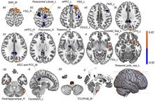
          A biomarker containing 3,650 features was identified with an accuracy of 79.3%, sensitivity of 78.6%, and specificity of 80.0% ( p < 0.002). The discriminative features were found in the trigeminal cervical complex/rostral ventromedial medulla (TCC/RVM), thalamus, medial prefrontal cortex (mPFC), and temporal gyrus. Then, 70 of 3,650 discriminative features were identified to predict the reduction of attack frequency after tVNS treatment with a correlation coefficient of 0.36 ( p = 0.03). The 70 predictive features were involved in TCC/RVM, mPFC, temporal gyrus, middle cingulate cortex (MCC), and insula. The reduction of migraine attack frequency had a positive correlation with right TCC/RVM (r = 0.433, p = 0.021), left MCC (r = 0.451, p = 0.016), and bilateral mPFC (r = 0.416, p = 0.028), and negative with left insula (r = −0.473, p = 0.011) and right superior temporal gyrus/middle temporal gyrus (r = −0.684, p < 0.001), respectively.

          Conclusions

          By machine learning, the study proposed two potential biomarkers that could discriminate patients with MWoA and predict the efficacy of tVNS in reducing migraine attack frequency. The pivotal features were mainly located in the TCC/RVM, thalamus, mPFC, and temporal gyrus.

          Related collections

          Most cited references87

          • Record: found
          • Abstract: not found
          • Article: not found

          Headache Classification Committee of the International Headache Society (IHS) The International Classification of Headache Disorders, 3rd edition

          (2018)
            Bookmark
            • Record: found
            • Abstract: found
            • Article: not found

            The brain's default network: anatomy, function, and relevance to disease.

            Thirty years of brain imaging research has converged to define the brain's default network-a novel and only recently appreciated brain system that participates in internal modes of cognition. Here we synthesize past observations to provide strong evidence that the default network is a specific, anatomically defined brain system preferentially active when individuals are not focused on the external environment. Analysis of connectional anatomy in the monkey supports the presence of an interconnected brain system. Providing insight into function, the default network is active when individuals are engaged in internally focused tasks including autobiographical memory retrieval, envisioning the future, and conceiving the perspectives of others. Probing the functional anatomy of the network in detail reveals that it is best understood as multiple interacting subsystems. The medial temporal lobe subsystem provides information from prior experiences in the form of memories and associations that are the building blocks of mental simulation. The medial prefrontal subsystem facilitates the flexible use of this information during the construction of self-relevant mental simulations. These two subsystems converge on important nodes of integration including the posterior cingulate cortex. The implications of these functional and anatomical observations are discussed in relation to possible adaptive roles of the default network for using past experiences to plan for the future, navigate social interactions, and maximize the utility of moments when we are not otherwise engaged by the external world. We conclude by discussing the relevance of the default network for understanding mental disorders including autism, schizophrenia, and Alzheimer's disease.
              Bookmark
              • Record: found
              • Abstract: found
              • Article: not found

              DPABI: Data Processing & Analysis for (Resting-State) Brain Imaging.

              Brain imaging efforts are being increasingly devoted to decode the functioning of the human brain. Among neuroimaging techniques, resting-state fMRI (R-fMRI) is currently expanding exponentially. Beyond the general neuroimaging analysis packages (e.g., SPM, AFNI and FSL), REST and DPARSF were developed to meet the increasing need of user-friendly toolboxes for R-fMRI data processing. To address recently identified methodological challenges of R-fMRI, we introduce the newly developed toolbox, DPABI, which was evolved from REST and DPARSF. DPABI incorporates recent research advances on head motion control and measurement standardization, thus allowing users to evaluate results using stringent control strategies. DPABI also emphasizes test-retest reliability and quality control of data processing. Furthermore, DPABI provides a user-friendly pipeline analysis toolkit for rat/monkey R-fMRI data analysis to reflect the rapid advances in animal imaging. In addition, DPABI includes preprocessing modules for task-based fMRI, voxel-based morphometry analysis, statistical analysis and results viewing. DPABI is designed to make data analysis require fewer manual operations, be less time-consuming, have a lower skill requirement, a smaller risk of inadvertent mistakes, and be more comparable across studies. We anticipate this open-source toolbox will assist novices and expert users alike and continue to support advancing R-fMRI methodology and its application to clinical translational studies.
                Bookmark

                Author and article information

                Contributors
                Journal
                Front Neurosci
                Front Neurosci
                Front. Neurosci.
                Frontiers in Neuroscience
                Frontiers Media S.A.
                1662-4548
                1662-453X
                05 August 2022
                2022
                : 16
                : 937453
                Affiliations
                [1] 1Department of Radiology, The Second Affiliated Hospital of Guangzhou University of Chinese Medicine , Guangzhou, China
                [2] 2The Second Clinical College, Guangzhou University of Chinese Medicine , Guangzhou, China
                [3] 3Department of Radiology, The First Affiliated Hospital of Henan University of Chinese Medicine , Zhengzhou, China
                Author notes

                Edited by: Mariella Pazzaglia, Sapienza University of Rome, Italy

                Reviewed by: Igor Petrusic, University of Belgrade, Serbia; Noemi Meylakh, The University of Sydney, Australia; Songhua Zhan, Affiliated to Shanghai University of Traditional Chinese Medicine, China

                *Correspondence: Bo Liu liubogzcm@ 123456163.com

                This article was submitted to Perception Science, a section of the journal Frontiers in Neuroscience

                †These authors share first authorship

                Article
                10.3389/fnins.2022.937453
                9388938
                35992927
                f3a9bb12-5609-4cc6-be81-b96eac6f2667
                Copyright © 2022 Fu, Zhang, Ye, Hou, Wen, Yan, Luo, Feng and Liu.

                This is an open-access article distributed under the terms of the Creative Commons Attribution License (CC BY). The use, distribution or reproduction in other forums is permitted, provided the original author(s) and the copyright owner(s) are credited and that the original publication in this journal is cited, in accordance with accepted academic practice. No use, distribution or reproduction is permitted which does not comply with these terms.

                History
                : 06 May 2022
                : 13 July 2022
                Page count
                Figures: 7, Tables: 3, Equations: 0, References: 87, Pages: 15, Words: 9101
                Funding
                Funded by: Guangzhou Municipal Science and Technology Project, doi 10.13039/501100010256;
                Funded by: Guangdong Provincial Hospital of Traditional Chinese Medicine, doi 10.13039/100012829;
                Categories
                Neuroscience
                Original Research

                Neurosciences
                migraine,transcutaneous vagus nerve stimulation,machine learning,functional magnetic resonance imaging,support vector machine (svm),vagus nerve

                Comments

                Comment on this article