35
views
0
recommends
+1 Recommend
0 collections
    0
    shares
      • Record: found
      • Abstract: found
      • Article: found
      Is Open Access

      Up-regulation of multiple proteins and biological processes during maxillary expansion in rats

      research-article

      Read this article at

      Bookmark
          There is no author summary for this article yet. Authors can add summaries to their articles on ScienceOpen to make them more accessible to a non-specialist audience.

          Abstract

          Background

          Maxillary expansion (ME) is a common practice in orthodontics that aims to increase the constricted maxillary arch width. Relapse often occurs, however, and better treatment strategies are needed. In order to develop a more effective method, this study was designed to further examine the process of tissue remodeling during ME, to identify the changes in expression of several proteins of interest, and to clarify the molecular mechanism responsible for tissue remodeling.

          Methods

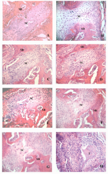
          Male Wistar rats were randomly divided into control and ME groups. The rats were euthanized at various intervals over 11 days, and the dissected palates were prepared for histological examination. The structure of the midpalatal sutures changed little during the first three days. Proteins from samples in the ground midpalatal tissues obtained on the third day were subjected to two-dimensional polyacrylamide gel electrophoresis (2-DE) and matrix assisted laser desorption/ionization-time of flight mass spectrometry (MALDI-TOF MS) analysis. Validation of protein expression was performed by Western blot analyses.

          Results

          From day 5, chondrocytes in the inner layer of suture cartilage and osteoblasts at the end of the suture cartilage began to proliferate, and the skeletal matrix increased later adjacent to the cartilage in the ME group. Comparative proteomic analysis showed increases in 22 protein spots present in the ME group. The changes in three proteins closely related to osteogenesis (parathyroid hormone, osteoprotegerin and vimentin) were confirmed by Western blotting.

          Conclusion

          Many proteins are over-expressed during ME, and they may play an important role in the remodeling process.

          Related collections

          Most cited references29

          • Record: found
          • Abstract: found
          • Article: not found

          Recombinant human parathyroid hormone (1-34) [teriparatide] improves both cortical and cancellous bone structure.

          Histomorphometry and microCT of 51 paired iliac crest biopsy specimens from women treated with teriparatide revealed significant increases in cancellous bone volume, cancellous bone connectivity density, cancellous bone plate-like structure, and cortical thickness, and a reduction in marrow star volume. We studied the ability of teriparatide (rDNA origin) injection [rhPTH(1-34), TPTD] to improve both cancellous and cortical bone in a subset of women enrolled in the Fracture Prevention Trial of postmenopausal women with osteoporosis after a mean treatment time of 19 months. This is the first report of a biopsy study after treatment with teriparatide having a sufficient number of paired biopsy samples to provide quantitative structural data. Fifty-one paired iliac crest bone biopsy specimens (placebo [n = 19], 20 microg teriparatide [n = 18], and 40 microg teriparatide [n = 14]) were analyzed using both two-dimensional (2D) histomorphometry and three-dimensional (3D) microcomputed tomography (microCT). Data for both teriparatide treatment groups were pooled for analysis. By 2D histomorphometric analyses, teriparatide significantly increased cancellous bone volume (median percent change: teriparatide, 14%; placebo, -24%; p = 0.001) and reduced marrow star volume (teriparatide, -16%; placebo, 112%; p = 0.004). Teriparatide administration was not associated with osteomalacia or woven bone, and there were no significant changes in mineral appositional rate or wall thickness. By 3D cancellous and cortical bone structural analyses, teriparatide significantly decreased the cancellous structure model index (teriparatide, -12%; placebo, 7%; p = 0.025), increased cancellous connectivity density (teriparatide, 19%; placebo, - 14%; p = 0.034), and increased cortical thickness (teriparatide, 22%; placebo, 3%; p = 0.012). These data show that teriparatide treatment of postmenopausal women with osteoporosis significantly increased cancellous bone volume and connectivity, improved trabecular morphology with a shift toward a more plate-like structure, and increased cortical bone thickness. These changes in cancellous and cortical bone morphology should improve biomechanical competence and are consistent with the substantially reduced incidences of vertebral and nonvertebral fractures during administration of teriparatide.
            Bookmark
            • Record: found
            • Abstract: found
            • Article: not found

            ENO1 gene product binds to the c-myc promoter and acts as a transcriptional repressor: relationship with Myc promoter-binding protein 1 (MBP-1).

            The Myc promoter-binding protein-1 (MBP-1) is a 37-38 kDa protein that binds to the c-myc P2 promoter and negatively regulates transcription of the protooncogene. MBP-1 cDNA shares 97% similarity with the cDNA encoding the glycolytic enzyme alpha-enolase and both genes have been mapped to the same region of human chromosome 1, suggesting the hypothesis that the two proteins might be encoded by the same gene. We show here data indicating that a 37 kDa protein is alternatively translated from the full-length alpha-enolase mRNA. This shorter form of alpha-enolase is able to bind the MBP-1 consensus sequence and to downregulate expression of a luciferase reporter gene under the control of the c-myc P2 promoter. Furthermore, using alpha-enolase/green fluorescent protein chimeras in transfection experiments we show that, while the 48 kDa alpha-enolase mainly has a cytoplasmic localization, the 37 kDa alpha-enolase is preferentially localized in the cell nuclei. The finding that a transcriptional repressor of the c-myc oncogene is an alternatively translated product of the ENO1 gene, which maps to a region of human chromosome 1 frequently deleted in human cancers, makes ENO1 a potential candidate for tumor suppressor.
              Bookmark
              • Record: found
              • Abstract: found
              • Article: not found

              New insights into the molecular mechanisms of action of bisphosphonates.

              Bisphosphonates are currently the most important and effective class of anti-resorptive drugs available, but the exact molecular mechanisms by which they inhibit osteoclast-mediated bone resorption have only recently been identified. Due to the targeting of bisphosphonates to bone mineral and the ability of osteoclasts to release bone-bound bisphosphonate, a direct effect on mature osteoclasts appears to be the most important route of action. As a result of recent discoveries concerning their molecular mechanism of action, bisphosphonates can be grouped into two classes. The simple bisphosphonates that closely resemble PPi (such as clodronate, etidronate and tiludronate) can be metabolically incorporated into non-hydrolysable analogues of ATP that accumulate intracellularly in osteoclasts, resulting in induction of osteoclast apoptosis. By contrast, the more potent, nitrogen-containing bisphosphonates (such as pamidronate, alendronate, risedronate, ibandronate and zoledronate) appear to act as analogues of isoprenoid diphosphate lipids, thereby inhibiting FPP synthase, an enzyme in the mevalonate pathway. Inhibition of this enzyme in osteoclasts prevents the biosynthesis of isoprenoid lipids (FPP and GGPP) that are essential for the post-translational farnesylation and geranylgeranylation of small GTPase signalling proteins. Loss of bone-resorptive activity and osteoclast apoptosis is due primarily to loss of geranylgeranylated small GTPases. Identification of FPP synthase as the target of nitrogen-containing bisphosphonates has also helped explain the molecular basis for the adverse effects of these agents in the GI tract and on the immune system.
                Bookmark

                Author and article information

                Journal
                BMC Musculoskelet Disord
                BMC Musculoskeletal Disorders
                BioMed Central
                1471-2474
                2008
                19 March 2008
                : 9
                : 37
                Affiliations
                [1 ]Institute of Stomatology, School of Stomatology, Nanjing Medical University, PR China
                [2 ]School of Dentistry, Faculty of Health Sciences, The University of Adelaide, Adelaide 5005, Australia
                Article
                1471-2474-9-37
                10.1186/1471-2474-9-37
                2291465
                18366685
                f2f5948d-d95c-45c1-9f09-19677dd5da19
                Copyright © 2008 Ma et al; licensee BioMed Central Ltd.

                This is an Open Access article distributed under the terms of the Creative Commons Attribution License ( http://creativecommons.org/licenses/by/2.0), which permits unrestricted use, distribution, and reproduction in any medium, provided the original work is properly cited.

                History
                : 22 July 2007
                : 19 March 2008
                Categories
                Research Article

                Orthopedics
                Orthopedics

                Comments

                Comment on this article