748
views
0
recommends
+1 Recommend
0 collections
    0
    shares
      • Record: found
      • Abstract: found
      • Article: found
      Is Open Access

      A gallery of the key characters to ease identification of Dermanyssus gallinae (Acari: Gamasida: Dermanyssidae) and allow differentiation from Ornithonyssus sylviarum (Acari: Gamasida: Macronyssidae)

      research-article

      Read this article at

      Bookmark
          There is no author summary for this article yet. Authors can add summaries to their articles on ScienceOpen to make them more accessible to a non-specialist audience.

          Abstract

          Background

          Dermanyssus gallinae (poultry red mite) is a major threat for the poultry industry and is of significant interest for public health. Identification of D. gallinae can be difficult for scientists not familiar with mite morphology and terminology especially when trying to use identification keys. Moreover, this species may easily be confused with another dermanyssoid mite, Ornithonyssus sylviarum (northern fowl mite), which often shares the same hosts and environment.

          Methods

          Specimens of D. gallinae were collected at poultry farms in the Puglia and performed for light and scanning electron microscopy observations, identification and micrographs. Moreover specimens of O. sylviarum were collected separately macerated and mounted on slides for light microscopy observations, identification and pictures.

          Results

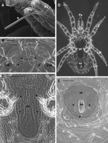
          The micrographs used in this study, based on LM and SEM observations, highlight the following important identifying characters of D. gallinae: the prominent shoulders of the dorsal shield and the jagged edges of the shield reticulations, the position of setae j1, s1 and the epigynal pores, and the presence on tibia IV pl of one seta. Additional micrographs highlighting the shape of the dorsal (abruptly narrowed posteriorly) and epigynal (narrowly rounded posteriorly) shields and the chelicera (elongate, with distinct digits) of O. sylviarum enable its differentiation from D.gallinae.

          Conclusion

          The photographic support provided here (both LM and SEM pictures) can be considered a practical tool for scientists who are not well acquainted with the morphology of D.gallinae, and who are involved with classical and molecular systematics, veterinary and human health aspects of poultry red mites.

          Related collections

          Most cited references17

          • Record: found
          • Abstract: not found
          • Article: not found

          Poultry integrated pest management: status and future

            Bookmark
            • Record: found
            • Abstract: found
            • Article: not found

            Vectorial role of some dermanyssoid mites (Acari, Mesostigmata, Dermanyssoidea).

            Among transmissible diseases, vectorial diseases represent a major problem for public health. In the group of acarina, while ticks are the most commonly implicated vectors, other arthropods and notably Dermanyssoidea are also involved in the transmission of pathogenic agents. Since the role of this superfamily is at present largely unknown, we have reviewed the vectorial role of these mites in the appearance, survival and propagation of pathogens. Various authors have shown that Dermanyssoidea are implicated in the transmission of both bacteria (Salmonella, Spirocheta, Rickettsia or Pasteurella) and viruses (equine encephalitis viruses, West Nile virus, Fowl pox virus, the virus causing Newcastle disease and tick borne encephalitis viruses or hantaviruses). Finally, some authors have also shown their role in the transmission of some protozoa and filaria. As the vectorial character of such mites has been more clearly demonstrated (Dermanyssus gallinae, Omithonyssus bacoti and Allodermanyssus sanguineus), it would be interesting to continue studies to better understand the role of this superfamily in the epidemiology of certain zoonoses.
              Bookmark
              • Record: found
              • Abstract: not found
              • Article: not found
              Is Open Access

              Beak condition and cage density determine abundance and spatial distribution of northern fowl mites, Ornithonyssus sylviarum, and chicken body lice, Menacanthus stramineus, on caged laying hens.

              Adult White Leghorn hens (Hy-Line strain W-36) were inoculated with either northern fowl mites or chicken body lice, and the ectoparasite populations were monitored over periods of 9 to 16 wk. Two beak conditions (beak trimmed or beak intact) and 2 housing densities (1 or 2 hens per 25 × 31 cm suspended wire cage) were tested. Populations of both ectoparasites were at least 10 times lower on beak-intact hens compared with populations on beak-trimmed hens. Cage density did not influence mite numbers, but higher numbers of lice (2 to 3 times) developed on hens held at the higher cage density. Louse distribution on the body and louse population age structure were also influenced by host beak condition. Beak-intact hens had a higher proportion of lice under the wings, whereas beak-trimmed hens had the majority of lice on the lower abdomen. Louse populations on beak-trimmed hens also comprised relatively more immature stages than populations found on beak-intact hens. The effects are likely related to decreased grooming efficiency by beak-trimmed hens and, in the case of lice, the higher host density. The high mite and louse populations on most commercial caged laying hens are probably a direct result of beak trimming. However, selection of more docile breeds that can be held without trimming may allow the hens themselves to reduce ectoparasites below economically damaging levels. This could benefit producers, animal welfare advocates, and human health by reducing 1) costs of beak trimming, 2) pesticide treatment costs (including human and bird chemical exposure concerns), and 3) objections to beak trimming from the animal welfare community.
                Bookmark

                Author and article information

                Journal
                Parasit Vectors
                Parasit Vectors
                Parasites & Vectors
                BioMed Central
                1756-3305
                2012
                30 May 2012
                : 5
                : 104
                Affiliations
                [1 ]Dipartimento di Scienze Agroambientali Chimica e Difesa Vegetale (DiSACD), University of Foggia, Via Napoli 25, 71100, Foggia, Italy
                [2 ]Dipartimento di Scienze delle Produzioni e dell'Innovazione nei Sistemi Agro-alimentari Mediterranei (PRIME), University of Foggia, Via Napoli 25, 71100, Foggia, Italy
                [3 ]Istituto Zooprofilattico Sperimentale della Puglia e della Basilicata, Via Manfredonia 20, 71100, Foggia, Italy
                Article
                1756-3305-5-104
                10.1186/1756-3305-5-104
                3419681
                22647594
                f21b0908-4cc5-43cc-80e5-3808370ee2ab
                Copyright ©2012 Di Palma et al.; licensee BioMed Central Ltd.

                This is an Open Access article distributed under the terms of the Creative Commons Attribution License ( http://creativecommons.org/licenses/by/2.0), which permits unrestricted use, distribution, and reproduction in any medium, provided the original work is properly cited.

                History
                : 4 January 2012
                : 6 April 2012
                Categories
                Research

                Parasitology
                poultry red mite,external morphology,northern fowl mite,identification,scanning electron microscopy,light microscopy

                Comments

                Comment on this article

                scite_
                95
                2
                61
                0
                Smart Citations
                95
                2
                61
                0
                Citing PublicationsSupportingMentioningContrasting
                View Citations

                See how this article has been cited at scite.ai

                scite shows how a scientific paper has been cited by providing the context of the citation, a classification describing whether it supports, mentions, or contrasts the cited claim, and a label indicating in which section the citation was made.

                Similar content175

                Cited by29

                Most referenced authors116