14
views
0
recommends
+1 Recommend
0 collections
    0
    shares
      • Record: found
      • Abstract: found
      • Article: found
      Is Open Access

      Toll-like Receptor Activation Induces Degeneration of Human Intervertebral Discs

      research-article

      Read this article at

      Bookmark
          There is no author summary for this article yet. Authors can add summaries to their articles on ScienceOpen to make them more accessible to a non-specialist audience.

          Abstract

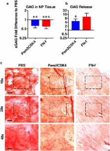
          Toll-like receptors (TLR) are activated by endogenous alarmins such as fragmented extracellular matrix compounds found in the degenerating disc. TLRs regulate cytokine, neurotrophin, and protease expression in human disc cells in vitro, and thus control key factors in disc degeneration. However, whether TLR activation leads to degenerative changes in intact human discs is unclear. Nucleus pulposus (NP) cells isolated from non-degenerating discs increase IL-1β and nerve growth factor gene expression following treatment with Pam2CSK4 (TLR2/6 agonist) but not Pam3CSK4 (TLR1/2 agonist). Challenging NP cells with Pam2CSK4 or 30 kDa fibronectin fragments (FN-f, an endogenous TLR2 and TLR4 alarmin) increased secretion of proinflammatory cytokines. We then investigated the effect of TLR activation in intact, non-degenerate, ex vivo human discs. Discs were injected with PBS, Pam2CSK4 and FN-f, and cultured for 28 days. TLR activation increased proteoglycan and ECM protein release into the culture media and decreased proteoglycan content in the NP. Proteases, including MMP3, 13 and HTRA1, are secreted at higher levels following TLR activation. In addition, proinflammatory cytokine levels, including IL-6, TNFα and IFNγ, increased following TLR activation. These results indicate that TLR activation induces degeneration in human discs. Therefore, TLRs are potential disease-modifying therapeutic targets to slow disc degeneration.

          Related collections

          Most cited references39

          • Record: found
          • Abstract: found
          • Article: not found

          Role of cytokines in intervertebral disc degeneration: pain and disc content.

          Degeneration of the intervertebral discs (IVDs) is a major contributor to back, neck and radicular pain. IVD degeneration is characterized by increases in levels of the proinflammatory cytokines TNF, IL-1α, IL-1β, IL-6 and IL-17 secreted by the IVD cells; these cytokines promote extracellular matrix degradation, chemokine production and changes in IVD cell phenotype. The resulting imbalance in catabolic and anabolic responses leads to the degeneration of IVD tissues, as well as disc herniation and radicular pain. The release of chemokines from degenerating discs promotes the infiltration and activation of immune cells, further amplifying the inflammatory cascade. Leukocyte migration into the IVD is accompanied by the appearance of microvasculature tissue and nerve fibres. Furthermore, neurogenic factors, generated by both disc and immune cells, induce expression of pain-associated cation channels in the dorsal root ganglion. Depolarization of these ion channels is likely to promote discogenic and radicular pain, and reinforce the cytokine-mediated degenerative cascade. Taken together, an enhanced understanding of the contribution of cytokines and immune cells to these catabolic, angiogenic and nociceptive processes could provide new targets for the treatment of symptomatic disc disease. In this Review, the role of key inflammatory cytokines during each of the individual phases of degenerative disc disease, as well as the outcomes of major clinical studies aimed at blocking cytokine function, are discussed.
            Bookmark
            • Record: found
            • Abstract: found
            • Article: not found

            What is intervertebral disc degeneration, and what causes it?

            Review and reinterpretation of existing literature. To suggest how intervertebral disc degeneration might be distinguished from the physiologic processes of growth, aging, healing, and adaptive remodeling. The research literature concerning disc degeneration is particularly diverse, and there are no accepted definitions to guide biomedical research, or medicolegal practice. The process of disc degeneration is an aberrant, cell-mediated response to progressive structural failure. A degenerate disc is one with structural failure combined with accelerated or advanced signs of aging. Early degenerative changes should refer to accelerated age-related changes in a structurally intact disc. Degenerative disc disease should be applied to a degenerate disc that is also painful. Structural defects such as endplate fracture, radial fissures, and herniation are easily detected, unambiguous markers of impaired disc function. They are not inevitable with age and are more closely related to pain than any other feature of aging discs. Structural failure is irreversible because adult discs have limited healing potential. It also progresses by physical and biologic mechanisms, and, therefore, is a suitable marker for a degenerative process. Biologic progression occurs because structural failure uncouples the local mechanical environment of disc cells from the overall loading of the disc, so that disc cell responses can be inappropriate or "aberrant." Animal models confirm that cell-mediated changes always follow structural failure caused by trauma. This definition of disc degeneration simplifies the issue of causality: excessive mechanical loading disrupts a disc's structure and precipitates a cascade of cell-mediated responses, leading to further disruption. Underlying causes of disc degeneration include genetic inheritance, age, inadequate metabolite transport, and loading history, all of which can weaken discs to such an extent that structural failure occurs during the activities of daily living. The other closely related definitions help to distinguish between degenerate and injured discs, and between discs that are and are not painful.
              Bookmark
              • Record: found
              • Abstract: found
              • Article: found
              Is Open Access

              Catabolic cytokine expression in degenerate and herniated human intervertebral discs: IL-1β and TNFα expression profile

              Low back pain is a common and debilitating disorder. Current evidence implicates intervertebral disc (IVD) degeneration and herniation as major causes, although the pathogenesis is poorly understood. While several cytokines have been implicated in the process of IVD degeneration and herniation, investigations have predominately focused on Interleukin 1 (IL-1) and tumor necrosis factor alpha (TNFα). However, to date no studies have investigated the expression of these cytokines simultaneously in IVD degeneration or herniation, or determined which may be the predominant cytokine associated with these disease states. Using quantitative real time PCR and immunohistochemistry we investigated gene and protein expression for IL-1β, TNFα and their receptors in non-degenerate, degenerate and herniated human IVDs. IL-1β gene expression was observed in a greater proportion of IVDs than TNFα (79% versus 59%). Degenerate and herniated IVDs displayed higher levels of both cytokines than non-degenerate IVDs, although in degenerate IVDs higher levels of IL-1β gene expression (1,300 copies/100 ng cDNA) were observed compared to those of TNFα (250 copies of TNFα/100 ng cDNA). Degenerate IVDs showed ten-fold higher IL-1 receptor gene expression compared to non-degenerate IVDs. In addition, 80% of degenerate IVD cells displayed IL-1 receptor immunopositivity compared to only 30% of cells in non-degenerate IVDs. However, no increase in TNF receptor I gene or protein expression was observed in degenerate or herniated IVDs compared to non-degenerate IVDs. We have demonstrated that although both cytokines are produced by human IVD cells, IL-1β is expressed at higher levels and in more IVDs, particularly in more degenerate IVDs (grades 4 to 12). Importantly, this study has highlighted an increase in gene and protein production for the IL-1 receptor type I but not the TNF receptor type I in degenerate IVDs. The data thus suggest that although both cytokines may be involved in the pathogenesis of IVD degeneration, IL-1 may have a more significant role than TNFα, and thus may be a better target for therapeutic intervention.
                Bookmark

                Author and article information

                Contributors
                lisbet.haglund@mcgill.ca
                Journal
                Sci Rep
                Sci Rep
                Scientific Reports
                Nature Publishing Group UK (London )
                2045-2322
                7 December 2017
                7 December 2017
                2017
                : 7
                : 17184
                Affiliations
                [1 ]ISNI 0000 0000 9064 4811, GRID grid.63984.30, Orthopaedic Research Lab, Department of Surgery, , McGill University and the Research Institute of the McGill University Health Centre, ; 1650 Cedar Ave, C10-148.8, Montreal, QC, H3G 1A4 Canada
                [2 ]ISNI 0000 0000 9064 4811, GRID grid.63984.30, McGill Scoliosis and Spine Group, Department of Surgery, , McGill University and the Research Institute of the McGill University Health Centre, ; 1650 Cedar Ave, C10-148.8, Montreal, QC, H3G 1A4 Canada
                [3 ]ISNI 0000 0004 0629 1363, GRID grid.415833.8, Shriner’s Hospital for Children, ; 1003 Decarie Blvd, Montreal, QC, H4A 0A9 Canada
                Author information
                http://orcid.org/0000-0002-8501-2945
                http://orcid.org/0000-0002-1288-2149
                Article
                17472
                10.1038/s41598-017-17472-1
                5719358
                29215065
                f20b087a-fcc2-4c79-aae4-d5372b3fa3c8
                © The Author(s) 2017

                Open Access This article is licensed under a Creative Commons Attribution 4.0 International License, which permits use, sharing, adaptation, distribution and reproduction in any medium or format, as long as you give appropriate credit to the original author(s) and the source, provide a link to the Creative Commons license, and indicate if changes were made. The images or other third party material in this article are included in the article’s Creative Commons license, unless indicated otherwise in a credit line to the material. If material is not included in the article’s Creative Commons license and your intended use is not permitted by statutory regulation or exceeds the permitted use, you will need to obtain permission directly from the copyright holder. To view a copy of this license, visit http://creativecommons.org/licenses/by/4.0/.

                History
                : 24 August 2017
                : 26 November 2017
                Categories
                Article
                Custom metadata
                © The Author(s) 2017

                Uncategorized
                Uncategorized

                Comments

                Comment on this article