11
views
0
recommends
+1 Recommend
0 collections
    0
    shares
      • Record: found
      • Abstract: found
      • Article: found
      Is Open Access

      Metabolic Features of Mouse and Human Retinas: Rods versus Cones, Macula versus Periphery, Retina versus RPE

      research-article

      Read this article at

      Bookmark
          There is no author summary for this article yet. Authors can add summaries to their articles on ScienceOpen to make them more accessible to a non-specialist audience.

          Summary

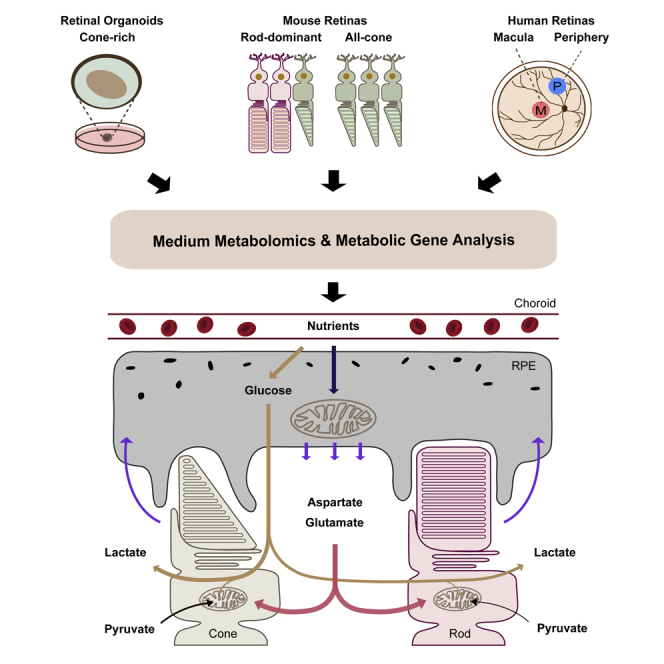
          Photoreceptors, especially cones, which are enriched in the human macula, have high energy demands, making them vulnerable to metabolic stress. Metabolic dysfunction of photoreceptors and their supporting retinal pigment epithelium (RPE) is an important underlying cause of degenerative retinal diseases. However, how cones and the macula support their exorbitant metabolic demand and communicate with RPE is unclear. By profiling metabolite uptake and release and analyzing metabolic genes, we have found cone-rich retinas and human macula share specific metabolic features with upregulated pathways in pyruvate metabolism, mitochondrial TCA cycle, and lipid synthesis. Human neural retina and RPE have distinct but complementary metabolic features. Retinal metabolism centers on NADH production and neurotransmitter biosynthesis. The retina needs aspartate to sustain its aerobic glycolysis and mitochondrial metabolism. RPE metabolism is directed toward NADPH production and biosynthesis of acetyl-rich metabolites, serine, and others. RPE consumes multiple nutrients, including proline, to produce metabolites for the retina.

          Graphical Abstract

          Highlights

          • Besides glucose, mouse and human retinas heavily consume aspartate and glutamate

          • Mature retinal organoids robustly consume aspartate and glutamate

          • Cone-rich retinas and the neural macula use more pyruvate

          • Neural retinas and RPE differ in their metabolite uptake and release

          Abstract

          Biochemistry; Sensory Neuroscience; Metabolomics

          Related collections

          Most cited references66

          • Record: found
          • Abstract: found
          • Article: not found

          Glucose feeds the TCA cycle via circulating lactate

          Mammalian tissues are fuelled by circulating nutrients, including glucose, amino acids, and various intermediary metabolites. Under aerobic conditions, glucose is generally assumed to be burned fully by tissues via the tricarboxylic acid cycle (TCA cycle) to carbon dioxide. Alternatively, glucose can be catabolized anaerobically via glycolysis to lactate, which is itself also a potential nutrient for tissues and tumours. The quantitative relevance of circulating lactate or other metabolic intermediates as fuels remains unclear. Here we systematically examine the fluxes of circulating metabolites in mice, and find that lactate can be a primary source of carbon for the TCA cycle and thus of energy. Intravenous infusions of 13C-labelled nutrients reveal that, on a molar basis, the circulatory turnover flux of lactate is the highest of all metabolites and exceeds that of glucose by 1.1-fold in fed mice and 2.5-fold in fasting mice; lactate is made primarily from glucose but also from other sources. In both fed and fasted mice, 13C-lactate extensively labels TCA cycle intermediates in all tissues. Quantitative analysis reveals that during the fasted state, the contribution of glucose to tissue TCA metabolism is primarily indirect (via circulating lactate) in all tissues except the brain. In genetically engineered lung and pancreatic cancer tumours in fasted mice, the contribution of circulating lactate to TCA cycle intermediates exceeds that of glucose, with glutamine making a larger contribution than lactate in pancreatic cancer. Thus, glycolysis and the TCA cycle are uncoupled at the level of lactate, which is a primary circulating TCA substrate in most tissues and tumours.
            Bookmark
            • Record: found
            • Abstract: found
            • Article: not found

            mTOR-mediated dedifferentiation of the retinal pigment epithelium initiates photoreceptor degeneration in mice.

            Retinal pigment epithelial (RPE) cell dysfunction plays a central role in various retinal degenerative diseases, but knowledge is limited regarding the pathways responsible for adult RPE stress responses in vivo. RPE mitochondrial dysfunction has been implicated in the pathogenesis of several forms of retinal degeneration. Here we have shown that postnatal ablation of RPE mitochondrial oxidative phosphorylation in mice triggers gradual epithelium dedifferentiation, typified by reduction of RPE-characteristic proteins and cellular hypertrophy. The electrical response of the retina to light decreased and photoreceptors eventually degenerated. Abnormal RPE cell behavior was associated with increased glycolysis and activation of, and dependence upon, the hepatocyte growth factor/met proto-oncogene pathway. RPE dedifferentiation and hypertrophy arose through stimulation of the AKT/mammalian target of rapamycin (AKT/mTOR) pathway. Administration of an oxidant to wild-type mice also caused RPE dedifferentiation and mTOR activation. Importantly, treatment with the mTOR inhibitor rapamycin blunted key aspects of dedifferentiation and preserved photoreceptor function for both insults. These results reveal an in vivo response of the mature RPE to diverse stressors that prolongs RPE cell survival at the expense of epithelial attributes and photoreceptor function. Our findings provide a rationale for mTOR pathway inhibition as a therapeutic strategy for retinal degenerative diseases involving RPE stress.
              Bookmark
              • Record: found
              • Abstract: found
              • Article: not found

              ATP consumption by mammalian rod photoreceptors in darkness and in light.

              Why do vertebrates use rods and cones that hyperpolarize, when in insect eyes a single depolarizing photoreceptor can function at all light levels? We answer this question at least in part with a comprehensive assessment of ATP consumption for mammalian rods from voltages and currents and recently published physiological and biochemical data. In darkness, rods consume 10(8) ATP s(-1), about the same as Drosophila photoreceptors. Ion fluxes associated with phototransduction and synaptic transmission dominate; as in CNS, the contribution of enzymes of the second-messenger cascade is surprisingly small. Suppression of rod responses in daylight closes light-gated channels and reduces total energy consumption by >75%, but in Drosophila light opens channels and increases consumption 5-fold. Rods therefore provide an energy-efficient mechanism not present in rhabdomeric photoreceptors. Rods are metabolically less "costly" than cones, because cones do not saturate in bright light and use more ATP s(-1) for transducin activation and rhodopsin phosphorylation. This helps to explain why the vertebrate retina is duplex, and why some diurnal animals like primates have a small number of cones, concentrated in a region of high acuity.
                Bookmark

                Author and article information

                Contributors
                Journal
                iScience
                iScience
                iScience
                Elsevier
                2589-0042
                14 October 2020
                20 November 2020
                14 October 2020
                : 23
                : 11
                : 101672
                Affiliations
                [1 ]Departments of Ophthalmology and Biochemistry, West Virginia University, WVU Eye Institute, One Medical Center Dr, PO Box 9193, Morgantown, WV 26506, USA
                [2 ]Department of Ophthalmology, Affiliated Hospital of Yangzhou University, Yangzhou University, Yangzhou, Jiangsu Province 225100, China
                [3 ]Save Sight Institute, Sydney Medical School, University of Sydney, Sydney, NSW 2000, Australia
                [4 ]Department of Ophthalmology and Visual Sciences, Albert Einstein College of Medicine, Bronx, NY 10461, USA
                [5 ]Department of Genetics, Albert Einstein College of Medicine, Bronx, NY 10461, USA
                Author notes
                []Corresponding author jianhai.du@ 123456wvumedicine.org
                [6]

                These authors contributed equally

                [7]

                Lead Contact

                Article
                S2589-0042(20)30864-6 101672
                10.1016/j.isci.2020.101672
                7644940
                33196018
                f185683c-9dcd-43a5-a3bf-23f5f7760deb
                © 2020 The Author(s)

                This is an open access article under the CC BY-NC-ND license (http://creativecommons.org/licenses/by-nc-nd/4.0/).

                History
                : 31 July 2020
                : 21 September 2020
                : 8 October 2020
                Categories
                Article

                biochemistry,sensory neuroscience,metabolomics
                biochemistry, sensory neuroscience, metabolomics

                Comments

                Comment on this article