255
views
0
recommends
+1 Recommend
0 collections
    0
    shares
      • Record: found
      • Abstract: found
      • Article: found
      Is Open Access

      A Nup133-dependent NPC-anchored network tethers centrosomes to the nuclear envelope in prophase

      research-article

      Read this article at

      Bookmark
          There is no author summary for this article yet. Authors can add summaries to their articles on ScienceOpen to make them more accessible to a non-specialist audience.

          Abstract

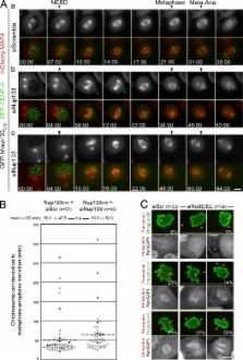
          Nup133 links CENP-F, NudE/EL, and the dynein/dynactin complex to anchor centrosomes to the nuclear membrane.

          Abstract

          Centrosomes are closely associated with the nuclear envelope (NE) throughout the cell cycle and this association is maintained in prophase when they separate to establish the future mitotic spindle. At this stage, the kinetochore constituents CENP-F, NudE, NudEL, dynein, and dynactin accumulate at the NE. We demonstrate here that the N-terminal domain of the nuclear pore complex (NPC) protein Nup133, although largely dispensable for NPC assembly, is required for efficient anchoring of the dynein/dynactin complex to the NE in prophase. Nup133 exerts this function through an interaction network via CENP-F and NudE/EL. We show that this molecular chain is critical for maintaining centrosome association with the NE at mitotic entry and contributes to this process without interfering with the previously described RanBP2–BICD2-dependent pathway of centrosome anchoring. Finally, our study reveals that tethering of centrosomes to the nuclear surface at the G2/M transition contributes, along with other cellular mechanisms, to early stages of bipolar spindle assembly.

          Related collections

          Most cited references52

          • Record: found
          • Abstract: found
          • Article: not found

          Small molecule inhibitor of mitotic spindle bipolarity identified in a phenotype-based screen.

          Small molecules that perturb specific protein functions are valuable tools for dissecting complex processes in mammalian cells. A combination of two phenotype-based screens, one based on a specific posttranslational modification, the other visualizing microtubules and chromatin, was used to identify compounds that affect mitosis. One compound, here named monastrol, arrested mammalian cells in mitosis with monopolar spindles. In vitro, monastrol specifically inhibited the motility of the mitotic kinesin Eg5, a motor protein required for spindle bipolarity. All previously known small molecules that specifically affect the mitotic machinery target tubulin. Monastrol will therefore be a particularly useful tool for studying mitotic mechanisms.
            Bookmark
            • Record: found
            • Abstract: found
            • Article: not found

            Visualization of microtubule growth in cultured neurons via the use of EB3-GFP (end-binding protein 3-green fluorescent protein).

            Several microtubule binding proteins, including CLIP-170 (cytoplasmic linker protein-170), CLIP-115, and EB1 (end-binding protein 1), have been shown to associate specifically with the ends of growing microtubules in non-neuronal cells, thereby regulating microtubule dynamics and the binding of microtubules to protein complexes, organelles, and membranes. When fused to GFP (green fluorescent protein), these proteins, which collectively are called +TIPs (plus end tracking proteins), also serve as powerful markers for visualizing microtubule growth events. Here we demonstrate that endogenous +TIPs are present at distal ends of microtubules in fixed neurons. Using EB3-GFP as a marker of microtubule growth in live cells, we subsequently analyze microtubule dynamics in neurons. Our results indicate that microtubules grow slower in neurons than in glia and COS-1 cells. The average speed and length of EB3-GFP movements are comparable in cell bodies, dendrites, axons, and growth cones. In the proximal region of differentiated dendrites approximately 65% of EB3-GFP movements are directed toward the distal end, whereas 35% are directed toward the cell body. In more distal dendritic regions and in axons most EB3-GFP dots move toward the growth cone. This difference in directionality of EB3-GFP movements in dendrites and axons reflects the highly specific microtubule organization in neurons. Together, these results suggest that local microtubule polymerization contributes to the formation of the microtubule network in all neuronal compartments. We propose that similar mechanisms underlie the specific association of CLIPs and EB1-related proteins with the ends of growing microtubules in non-neuronal and neuronal cells.
              Bookmark
              • Record: found
              • Abstract: found
              • Article: not found

              A general amphipathic alpha-helical motif for sensing membrane curvature.

              The Golgi-associated protein ArfGAP1 has an unusual membrane-adsorbing amphipathic alpha-helix: its polar face is weakly charged, containing mainly serine and threonine residues. We show that this feature explains the specificity of ArfGAP1 for curved versus flat lipid membranes. We built an algorithm to identify other potential amphipathic alpha-helices rich in serine and threonine residues in protein databases. Among the identified sequences, we show that three act as membrane curvature sensors. In the golgin GMAP-210, the sensor may serve to trap small vesicles at the end of a long coiled coil. In Osh4p/Kes1p, which transports sterol between membranes, the sensor controls access to the sterol-binding pocket. In the nucleoporin Nup133, the sensor corresponds to an exposed loop of a beta-propeller structure. Ser/Thr-rich amphipathic helices thus define a general motif used by proteins of various functions for sensing membrane curvature.
                Bookmark

                Author and article information

                Journal
                J Cell Biol
                J. Cell Biol
                jcb
                The Journal of Cell Biology
                The Rockefeller University Press
                0021-9525
                1540-8140
                7 March 2011
                : 192
                : 5
                : 855-871
                Affiliations
                [1 ]Cell Biology Program, Institut Jacques Monod, UMR 7592 Centre National de la Recherche Scientifique-Université Paris Diderot, 75205 Paris Cedex 13, France
                [2 ]Cell Biology and Biophysics Unit, European Molecular Biology Laboratory, D-69117 Heidelberg, Germany
                [3 ]Department of Pathology and Cell Biology, Columbia University, New York, NY 10032
                Author notes
                Correspondence to Valérie Doye: doye.valerie@ 123456ijm.univ-paris-diderot.fr

                S.Bolhy, I. Bouhlel, and E. Dultz contributed equally to this paper.

                Article
                201007118
                10.1083/jcb.201007118
                3051818
                21383080
                efc2ed9d-eae7-418d-a267-b30ae3e9a64b
                © 2011 Bolhy et al.

                This article is distributed under the terms of an Attribution–Noncommercial–Share Alike–No Mirror Sites license for the first six months after the publication date (see http://www.rupress.org/terms). After six months it is available under a Creative Commons License (Attribution–Noncommercial–Share Alike 3.0 Unported license, as described at http://creativecommons.org/licenses/by-nc-sa/3.0/).

                History
                : 21 July 2010
                : 9 February 2011
                Categories
                Research Articles
                Article

                Cell biology
                Cell biology

                Comments

                Comment on this article