6
views
0
recommends
+1 Recommend
0 collections
    0
    shares
      • Record: found
      • Abstract: found
      • Article: found
      Is Open Access

      Multiple comorbid neuropathologies in the setting of Alzheimer's disease neuropathology and implications for drug development

      review-article

      Read this article at

      Bookmark
          There is no author summary for this article yet. Authors can add summaries to their articles on ScienceOpen to make them more accessible to a non-specialist audience.

          Abstract

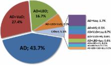
          Dementia is often characterized as being caused by one of several major diseases, such as Alzheimer's disease (AD), cerebrovascular disease, Lewy body disease, or a frontotemporal degeneration. Failure to acknowledge that more than one entity may be present precludes attempts to understand interactive relationships. The clinicopathological studies of dementia demonstrate that multiple pathologic processes often coexist.

          How overlapping pathologic findings affect the diagnosis and treatment of clinical AD and other dementia phenotypes was the topic taken up by the Alzheimer's Association's Research Roundtable in October 2014. This review will cover the neuropathologic basis of dementia, provide clinical perspectives on multiple pathologies, and discuss therapeutics and biomarkers targeting overlapping pathologies and how these issues impact clinical trials.High prevalence of multiple pathologic findings among individuals with clinical diagnosis of AD suggests that new treatment strategies may be needed to effectively treat AD and other dementing illnesses.

          Related collections

          Most cited references34

          • Record: found
          • Abstract: found
          • Article: not found

          Staging TDP-43 pathology in Alzheimer's disease.

          TDP-43 immunoreactivity occurs in 19-57 % of Alzheimer's disease (AD) cases. Two patterns of TDP-43 deposition in AD have been described involving hippocampus (limbic) or hippocampus and neocortex (diffuse), although focal amygdala involvement has been observed. In 195 AD cases with TDP-43, we investigated regional TDP-43 immunoreactivity with the aim of developing a TDP-43 in AD staging scheme. TDP-43 immunoreactivity was assessed in amygdala, entorhinal cortex, subiculum, hippocampal dentate gyrus, occipitotemporal, inferior temporal and frontal cortices, and basal ganglia. Clinical, neuroimaging, genetic and pathological characteristics were assessed across stages. Five stages were identified: stage I showed scant-sparse TDP-43 in the amygdala only (17 %); stage II showed moderate-frequent amygdala TDP-43 with spread into entorhinal and subiculum (25 %); stage III showed further spread into dentate gyrus and occipitotemporal cortex (31 %); stage IV showed further spread into inferior temporal cortex (20 %); and stage V showed involvement of frontal cortex and basal ganglia (7 %). Cognition and medial temporal volumes differed across all stages and progression across stages correlated with worsening cognition and medial temporal volume loss. Compared to 147 AD patients without TDP-43, only the Boston Naming Test showed abnormalities in stage I. The findings demonstrate that TDP-43 deposition in AD progresses in a stereotypic manner that can be divided into five distinct topographic stages which are supported by correlations with clinical and neuroimaging features. Given these findings, we recommend sequential regional TDP-43 screening in AD beginning with the amygdala.
            Bookmark
            • Record: found
            • Abstract: found
            • Article: not found

            Hippocampal sclerosis and TDP-43 pathology in aging and Alzheimer disease.

            To investigate the association of hippocampal sclerosis (HS) with TAR-DNA binding protein of 43kDa (TDP-43) and other common age-related pathologies, dementia, probable Alzheimer disease (AD), mild cognitive impairment (MCI), and cognitive domains in community-dwelling older subjects.
              Bookmark
              • Record: found
              • Abstract: found
              • Article: not found

              Cerebral microinfarcts: the invisible lesions.

              The association between small but still visible lacunar infarcts and cognitive decline has been established by population-based radiological and pathological studies. Microscopic examination of brain sections shows even smaller but substantially more numerous microinfarcts, the focus of this Review. These lesions often result from small vessel pathologies such as arteriolosclerosis or cerebral amyloid angiopathy. They typically go undetected in clinical-radiological correlation studies that rely on conventional structural MRI, although the largest acute microinfarcts can be detected by diffusion-weighted imaging. In view of their high numbers and widespread distribution, microinfarcts could directly disrupt important cognitive networks and thus account for some of the neurological dysfunction associated with lesions visible on conventional MRI such as lacunar infarcts and white matter hyperintensities. Standardised neuropathological assessment criteria and the development of non-invasive means of detection during life would be major steps towards understanding the causes and consequences of otherwise macroscopically invisible microinfarcts. Copyright © 2012 Elsevier Ltd. All rights reserved.
                Bookmark

                Author and article information

                Contributors
                Journal
                Alzheimers Dement (N Y)
                Alzheimers Dement (N Y)
                Alzheimer's & Dementia : Translational Research & Clinical Interventions
                Elsevier
                2352-8737
                20 September 2016
                January 2017
                20 September 2016
                : 3
                : 1
                : 83-91
                Affiliations
                [a ]Memory & Aging Center, Department of Neurology, University of California, San Francisco, San Francisco, CA, USA
                [b ]Division of Medical & Scientific Relations, Alzheimer's Association, Chicago IL, USA
                [c ]Merck, Kenilworth, NJ, USA
                [d ]Piramal Pharma, Inc., Boston, MA, USA
                [e ]Bracket Global, Wayne, PA, USA
                [f ]Quintiles (formerly), Rockville, MD, USA
                [g ]Department of Neurology, Mayo Clinic and Foundation, Rochester, MN, USA
                [h ]MedAvante, Hamilton, NJ, USA
                [i ]Department of Neurology, Loyola University Medical Center, Maywood, IL, USA
                [j ]Department of Clinical Neurosciences, Hotchkiss Brain Institute, University of Calgary, Calgary, Alberta, Canada
                [k ]European Medicines Agency (EMA), London, UK
                [l ]Clinical Memory Research Unit, Department of Clinical Sciences Malmö, Lund University, Malmö, Sweden
                [m ]Department of Neurology, Skåne University Hospital, Lund, Sweden
                [n ]Independent Science Writer, Elverson, PA, USA
                [o ]Eli Lilly & Co., Indianapolis, IN, USA
                Author notes
                []Corresponding author. Tel.: 312-604-1650; Fax: 877-444-6282. jhendrix@ 123456alz.org
                Article
                S2352-8737(16)30037-3
                10.1016/j.trci.2016.09.002
                5651346
                29067320
                ee884a63-7a16-4073-8d61-2ab9cf64c7a2
                © 2016 The Authors

                This is an open access article under the CC BY-NC-ND license (http://creativecommons.org/licenses/by-nc-nd/4.0/).

                History
                Categories
                Perspective

                alzheimer's disease (ad),cerebrovascular disease,lewy body disease,frontotemporal degeneration,β-amyloid,tau,α-synuclein,tdp-43

                Comments

                Comment on this article