16
views
0
recommends
+1 Recommend
0 collections
    0
    shares
      • Record: found
      • Abstract: found
      • Article: found
      Is Open Access

      Spred2 Deficiency Exacerbates D-Galactosamine/Lipopolysaccharide -induced Acute Liver Injury in Mice via Increased Production of TNFα

      research-article

      Read this article at

      Bookmark
          There is no author summary for this article yet. Authors can add summaries to their articles on ScienceOpen to make them more accessible to a non-specialist audience.

          Abstract

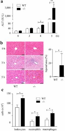
          Acute liver injury (ALI) is characterized by hepatocyte damage and inflammation. In the present study, we examined whether the absence of Sprouty-related EVH1-domain-containing protein 2 (Spred2), a negative regulator of the Ras/Raf/ERK/MAPK pathway, influences ALI induced by D-galactosamine (D-GalN) and lipopolysaccharide (LPS). Compared to wild-type mice, Spred2 −/− mice developed exacerbated liver injury represented by enhanced hepatocyte damage and inflammation. Enhanced ERK activation was observed in Spred2 −/−-livers, and the MEK/ERK inhibitor U0126 ameliorated ALI. Hepatic tumour necrosis factor α (TNFα) and interleukin (IL)-1β levels were increased in Spred-2 −/−-livers, and the neutralization of TNFα dramatically ameliorated ALI, which was associated with decreased levels of endogenous TNFα and IL-1β. When mice were challenged with D-GalN and TNFα, much severer ALI was observed in Spred2 −/− mice with significant increases in endogenous TNFα and IL-1β in the livers. Immunohistochemically, Kupffer cells were found to produce TNFα, and isolated Kupffer cells from Spred2 −/− mice produced significantly higher levels of TNFα than those from wild-type mice after LPS stimulation, which was significantly decreased by U0126. These results suggest that Spred2 negatively regulates D-GalN/LPS-induced ALI under the control of TNFα in Kupffer cells. Spred2 may present a therapeutic target for the treatment of ALI.

          Related collections

          Most cited references34

          • Record: found
          • Abstract: found
          • Article: not found

          Mitogen-activated protein kinases in innate immunity.

          Following pathogen infection or tissue damage, the stimulation of pattern recognition receptors on the cell surface and in the cytoplasm of innate immune cells activates members of each of the major mitogen-activated protein kinase (MAPK) subfamilies--the extracellular signal-regulated kinase (ERK), p38 and Jun N-terminal kinase (JNK) subfamilies. In conjunction with the activation of nuclear factor-κB and interferon-regulatory factor transcription factors, MAPK activation induces the expression of multiple genes that together regulate the inflammatory response. In this Review, we discuss our current knowledge about the regulation and the function of MAPKs in innate immunity, as well as the importance of negative feedback loops in limiting MAPK activity to prevent host tissue damage. We also examine how pathogens have evolved complex mechanisms to manipulate MAPK activation to increase their virulence. Finally, we consider the potential of the pharmacological targeting of MAPK pathways to treat autoimmune and inflammatory diseases.
            Bookmark
            • Record: found
            • Abstract: found
            • Article: not found

            Identification of a novel inhibitor of mitogen-activated protein kinase kinase.

            The compound U0126 (1,4-diamino-2,3-dicyano-1, 4-bis[2-aminophenylthio]butadiene) was identified as an inhibitor of AP-1 transactivation in a cell-based reporter assay. U0126 was also shown to inhibit endogenous promoters containing AP-1 response elements but did not affect genes lacking an AP-1 response element in their promoters. These effects of U0126 result from direct inhibition of the mitogen-activated protein kinase kinase family members, MEK-1 and MEK-2. Inhibition is selective for MEK-1 and -2, as U0126 shows little, if any, effect on the kinase activities of protein kinase C, Abl, Raf, MEKK, ERK, JNK, MKK-3, MKK-4/SEK, MKK-6, Cdk2, or Cdk4. Comparative kinetic analysis of U0126 and the MEK inhibitor PD098059 (Dudley, D. T., Pang, L., Decker, S. J., Bridges, A. J., and Saltiel, A. R. (1995) Proc. Natl. Acad. Sci U. S. A. 92, 7686-7689) demonstrates that U0126 and PD098059 are noncompetitive inhibitors with respect to both MEK substrates, ATP and ERK. We further demonstrate that the two compounds bind to deltaN3-S218E/S222D MEK in a mutually exclusive fashion, suggesting that they may share a common or overlapping binding site(s). Quantitative evaluation of the steady state kinetics of MEK inhibition by these compounds reveals that U0126 has approximately 100-fold higher affinity for deltaN3-S218E/S222D MEK than does PD098059. We further tested the effects of these compounds on the activity of wild type MEK isolated after activation from stimulated cells. Surprisingly, we observe a significant diminution in affinity of both compounds for wild type MEK as compared with the deltaN3-S218E/S222D mutant enzyme. These results suggest that the affinity of both compounds is mediated by subtle conformational differences between the two activated MEK forms. The MEK affinity of U0126, its selectivity for MEK over other kinases, and its cellular efficacy suggest that this compound will serve as a powerful tool for in vitro and cellular investigations of mitogen-activated protein kinase-mediated signal transduction.
              Bookmark
              • Record: found
              • Abstract: not found
              • Article: not found

              Preparation of isolated rat liver cells.

              P O Seglen (1976)
                Bookmark

                Author and article information

                Contributors
                amatsu@md.okayama-u.ac.jp
                Journal
                Sci Rep
                Sci Rep
                Scientific Reports
                Nature Publishing Group UK (London )
                2045-2322
                9 January 2018
                9 January 2018
                2018
                : 8
                : 188
                Affiliations
                [1 ]ISNI 0000 0001 1302 4472, GRID grid.261356.5, Department of Pathology and Experimental Medicine, , Graduate School of Medicine, Dentistry and Pharmaceutical Sciences, Okayama University, ; Okayama, Japan
                [2 ]ISNI 0000000086837370, GRID grid.214458.e, Department of Pathology, , University of Michigan Medical School, ; Ann Arbor, Michigan USA
                Article
                18380
                10.1038/s41598-017-18380-0
                5760641
                29317674
                ee630eb1-5cff-4563-9227-61de4af6b463
                © The Author(s) 2017

                Open Access This article is licensed under a Creative Commons Attribution 4.0 International License, which permits use, sharing, adaptation, distribution and reproduction in any medium or format, as long as you give appropriate credit to the original author(s) and the source, provide a link to the Creative Commons license, and indicate if changes were made. The images or other third party material in this article are included in the article’s Creative Commons license, unless indicated otherwise in a credit line to the material. If material is not included in the article’s Creative Commons license and your intended use is not permitted by statutory regulation or exceeds the permitted use, you will need to obtain permission directly from the copyright holder. To view a copy of this license, visit http://creativecommons.org/licenses/by/4.0/.

                History
                : 2 November 2017
                : 8 December 2017
                Categories
                Article
                Custom metadata
                © The Author(s) 2018

                Uncategorized
                Uncategorized

                Comments

                Comment on this article