12
views
0
recommends
+1 Recommend
0 collections
    0
    shares
      • Record: found
      • Abstract: found
      • Article: found
      Is Open Access

      Intravascular Papillary Endothelial Hyperplasia (Masson’s Tumor): Diagnosis the Plastic Surgeon Should Be Aware of

      case-report
      , MD * , , BS , , MD, PhD , , MD * ,
      Plastic and Reconstructive Surgery Global Open
      Wolters Kluwer Health

      Read this article at

      Bookmark
          There is no author summary for this article yet. Authors can add summaries to their articles on ScienceOpen to make them more accessible to a non-specialist audience.

          Summary:

          Intravascular papillary endothelial hyperplasia (IPEH) or Masson’s tumor is a rare benign entity commonly found on the head, neck, and upper extremities. It usually arises within a blood vessel but is considered to be a nonneoplastic reactive process often associated with vascular injury. Typically, IPEHs cause no symptoms and present as slowly growing soft-tissue masses. Given their prevalent location and indolent clinical presentation, the plastic surgeon should be familiar with this rare entity. We are presenting a case of IPEH of the forehead with unusual clinical and pathologic characteristics. Differential diagnosis, special considerations regarding preoperative work-up, and treatment options are discussed.

          Related collections

          Most cited references10

          • Record: found
          • Abstract: found
          • Article: not found

          Intravascular papillary endothelial hyperplasia. A clinicopathologic study of 91 cases.

          Ninety-one cases of intravascular papillary endothelial hyperplasia were studied clinically and histologically. This peculiar benign process, occasionally resembling hemangiosarcoma, was subgrouped in the following categories: a pure form that occurs within a dilated vascular space (30 cases), a mixed form that appears as a focal change in a hemangioma (55 cases), a third form (six cases) that belongs to neither of the first two. In the pure form, the lesions were most frequently situated in the subcutis of fingers (14 cases), of the head and neck (seven cases), and in the region between the elbows and hands (six cases). In the mixed form, half of the accompanying hemangiomas were intramuscular in no particular predilective sites. Papillary proliferation of endothelial cells was commonly found to be closely associated with thrombotic material and seemed to be an unusual feature of a thrombus undergoing organization.
            Bookmark
            • Record: found
            • Abstract: not found
            • Article: not found

            Hemangioendotheliome vegetant intravasculaire

              Bookmark
              • Record: found
              • Abstract: found
              • Article: found
              Is Open Access

              Intravascular papillary endothelial hyperplasia: histomorphological and immunohistochemical features

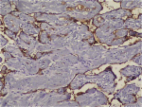
              Background Intravascular papillary endothelial hyperplasia (IPEH) is a benign intravascular process with features mimicking other benign and malignant vascular proliferations. IPEH lesions predominate in the head-neck region and the extremities. The characteristic histomorphological feature of IPEH is a papillary structure covered with hyperplastic endothelial cells within the vascular lumen. It is critical that this clinically benign lesion should not be mistaken for well-differentiated vascular tumors. In addition to the characteristic histological features, other useful diagnostic features included the intra-luminal location of the lesion, an intimate association with the organizing thrombus, the absence of necrosis, cellular pleomorphism, and mitotic activity. In addition, immunohistochemistry may indicate the vascular origin and proliferative index. In this study, we evaluated histomorphological and immunohistochemical findings (CD31, CD34, FVIII, type IV collagen, SMA, MSA, CD105, and Ki-67 staining) of ten IPEH cases. Methods Ten IPEH cases were re-examined for a panel of histomorphological and immunohistochemical features. CD31, CD34, FVIII, Type IV collagen, SMA and MSA antibodies utilized for immunohistochemical analysis. The histomorphological and immunohistochemical findings were evaluated by two independent pathologists using light microscopy. Results All ten cases involved intraluminal lesions with characteristic features of IPEH. All ten cases (100%) were stained positive for CD31 and CD34. The degree of staining with FVIII, type IV collagen, SMA, and MSA was variable. Conclusion In this series of specimens, CD31 and CD34 were the most sensitive markers indicating the vascular origin of the lesion. Staining for the other vascular markers (FVIII, type IV collagen, SMA and MSA) was variable. Different maturation degrees of lesions may account for the variation in immunohistochemical staining. Few previous investigations evaluated a wide range of antigen panels in IPEH sections. In our opinion, the evaluation of immune markers in a larger sample set will reveal new features in the maturity and developmental pathogenesis of vascular lesions and angiogenesis. IPEH is a benign lesion, which must be differentiated from malignant tumors such as angiosarcoma and Kaposi’s sarcoma. Improved definition of IPEH lesions using immunohistochemical markers may enhance the ability to differentiate between various vascular lesions. Virtual slides The virtual slide(s) for this article can be found here: http://www.diagnosticpathology.diagnomx.eu/vs/1381849312101856.
                Bookmark

                Author and article information

                Journal
                Plast Reconstr Surg Glob Open
                Plast Reconstr Surg Glob Open
                GOX
                Plastic and Reconstructive Surgery Global Open
                Wolters Kluwer Health
                2169-7574
                January 2017
                10 January 2017
                : 5
                : 1
                : e1122
                Affiliations
                From the [* ]Division of Plastic Surgery, Department of Surgery, University of Texas Medical Branch, Galveston, Tex.; []School of Medicine, University of Texas Medical Branch, Galveston, Tex.; and []Department of Surgical Pathology, University of Texas Medical Branch, Galveston, Tex.
                Author notes
                Eric Lowry Cole, MD, Division of Plastic Surgery, Department of Surgery, University of Texas Medical Branch, 301 University Blvd., Galveston, TX 77555, E-mail: elcole@ 123456utmb.edu
                Article
                00018
                10.1097/GOX.0000000000001122
                5293289
                28203491
                ed957fc0-1379-4e84-8676-30436a08e7f8
                Copyright © 2017 The Authors. Published by Wolters Kluwer Health, Inc. on behalf of The American Society of Plastic Surgeons. All rights reserved.

                This is an open-access article distributed under the terms of the Creative Commons Attribution-Non Commercial-No Derivatives License 4.0 (CCBY-NC-ND), where it is permissible to download and share the work provided it is properly cited. The work cannot be changed in any way or used commercially without permission from the journal.

                History
                : 23 May 2016
                : 16 September 2016
                Categories
                Case Report
                Custom metadata
                TRUE

                Comments

                Comment on this article