2
views
0
recommends
+1 Recommend
0 collections
    0
    shares
      • Record: found
      • Abstract: found
      • Article: found
      Is Open Access

      Fixation-induced surgical segment’s high stiffness and the damage of posterior structures together trigger a higher risk of adjacent segment disease in patients with lumbar interbody fusion operations

      research-article

      Read this article at

      Bookmark
          There is no author summary for this article yet. Authors can add summaries to their articles on ScienceOpen to make them more accessible to a non-specialist audience.

          Abstract

          Background

          Adjacent segment disease (ASD) is a commonly reported complication after lumbar interbody fusion (LIF); changes in the mechanical environment play an essential role in the generation of ASD. Traditionally, fixation-induced high stiffness in the surgical segment was the main reason for ASD. However, with more attention paid to the biomechanical significance of posterior bony and soft structures, surgeons hypothesize that this factor may also play an important role in ASD.

          Methods

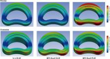
          Oblique and posterior LIF operations have been simulated in this study. The stand-alone OLIF and OLIF fixed by bilateral pedicle screw (BPS) system have been simulated. The spinal process (the attachment point of cranial ligamentum complex) was excised in the PLIF model; the BPS system has also been used in the PLIF model. Stress values related to ASD have been computed under physiological body positions, including flexion, extension, bending, and axial rotations.

          Results

          Compared to the stand-alone OLIF model, the OLIF model with BPS fixation suffers higher stress values under extension body position. However, there are no apparent differences under other loading conditions. Moreover, significant increases in stress values can be recorded in flexion and extension loading conditions in the PLIF model with posterior structures damage.

          Conclusions

          Fixation-induced surgical segment’s high stiffness and the damage of posterior soft tissues together trigger a higher risk of ASD in patients with LIF operations. Optimizing BPS fixation methods and pedicle screw designs and reducing the range of posterior structures excision may be an effective method to reduce the risk of ASD.

          Related collections

          Most cited references56

          • Record: found
          • Abstract: found
          • Article: not found

          Magnetic resonance classification of lumbar intervertebral disc degeneration.

          A reliability study was conducted. To develop a classification system for lumbar disc degeneration based on routine magnetic resonance imaging, to investigate the applicability of a simple algorithm, and to assess the reliability of this classification system. A standardized nomenclature in the assessment of disc abnormalities is a prerequisite for a comparison of data from different investigations. The reliability of the assessment has a crucial influence on the validity of the data. Grading systems of disc degeneration based on state of the art magnetic resonance imaging and corresponding reproducibility studies currently are sparse. A grading system for lumbar disc degeneration was developed on the basis of the literature. An algorithm to assess the grading was developed and optimized by reviewing lumbar magnetic resonance examinations. The reliability of the algorithm in depicting intervertebral disc alterations was tested on the magnetic resonance images of 300 lumbar intervertebral discs in 60 patients (33 men and 27 women) with a mean age of 40 years (range, 10-83 years). All scans were analyzed independently by three observers. Intra- and interobserver reliabilities were assessed by calculating kappa statistics. There were 14 Grade I, 82 Grade II, 72 Grade III, 68 Grade IV, and 64 Grade V discs. The kappa coefficients for intra- and interobserver agreement were substantial to excellent: intraobserver (kappa range, 0.84-0.90) and interobserver (kappa range, 0.69-0.81). Complete agreement was obtained, on the average, in 83.8% of all the discs. A difference of one grade occurred in 15.9% and a difference of two or more grades in 1.3% of all the cases. Disc degeneration can be graded reliably on routine T2-weighted magnetic resonance images using the grading system and algorithm presented in this investigation.
            Bookmark
            • Record: found
            • Abstract: found
            • Article: not found

            What is intervertebral disc degeneration, and what causes it?

            Review and reinterpretation of existing literature. To suggest how intervertebral disc degeneration might be distinguished from the physiologic processes of growth, aging, healing, and adaptive remodeling. The research literature concerning disc degeneration is particularly diverse, and there are no accepted definitions to guide biomedical research, or medicolegal practice. The process of disc degeneration is an aberrant, cell-mediated response to progressive structural failure. A degenerate disc is one with structural failure combined with accelerated or advanced signs of aging. Early degenerative changes should refer to accelerated age-related changes in a structurally intact disc. Degenerative disc disease should be applied to a degenerate disc that is also painful. Structural defects such as endplate fracture, radial fissures, and herniation are easily detected, unambiguous markers of impaired disc function. They are not inevitable with age and are more closely related to pain than any other feature of aging discs. Structural failure is irreversible because adult discs have limited healing potential. It also progresses by physical and biologic mechanisms, and, therefore, is a suitable marker for a degenerative process. Biologic progression occurs because structural failure uncouples the local mechanical environment of disc cells from the overall loading of the disc, so that disc cell responses can be inappropriate or "aberrant." Animal models confirm that cell-mediated changes always follow structural failure caused by trauma. This definition of disc degeneration simplifies the issue of causality: excessive mechanical loading disrupts a disc's structure and precipitates a cascade of cell-mediated responses, leading to further disruption. Underlying causes of disc degeneration include genetic inheritance, age, inadequate metabolite transport, and loading history, all of which can weaken discs to such an extent that structural failure occurs during the activities of daily living. The other closely related definitions help to distinguish between degenerate and injured discs, and between discs that are and are not painful.
              Bookmark
              • Record: found
              • Abstract: found
              • Article: not found

              Adjacent segment disease after lumbar or lumbosacral fusion: review of the literature.

              Review of the literature. Review the definition, etiology, incidence, and risk factors associated with as well as potential treatment options. The development of pathology at the mobile segment next to a lumbar or lumbosacral spinal fusion has been termed adjacent segment disease. Initially reported to occur rarely, it is now considered a potential late complication of spinal fusion that can necessitate further surgical intervention and adversely affect outcomes. MEDLINE literature search. The most common abnormal finding at the adjacent segment is disc degeneration. Biomechanical changes consisting of increased intradiscal pressure, increased facet loading, and increased mobility occur after fusion and have been implicated in causing adjacent segment disease. Progressive spinal degeneration with age is also thought to be a major contributor. From a radiographic standpoint, reported incidence during average postoperative follow-up observation ranging from 36 to 369 months varies substantially from 5.2 to 100%. Incidence of symptomatic adjacent segment disease is lower, however, ranging from 5.2 to 18.5% during 44.8 to 164 months of follow-up observation. The rate of symptomatic adjacent segment disease is higher in patients with transpedicular instrumentation (12.2-18.5%) compared with patients fused with other forms of instrumentation or with no instrumentation (5.2-5.6%). Potential risk factors include instrumentation, fusion length, sagittal malalignment, facet injury, age, and pre-existing degenerative changes. Biomechanical alterations likely play a primary role in causing adjacent segment disease. Radiographically apparent, asymptomatic adjacent segment disease is common but does not correlate with functional outcomes. Potentially modifiable risk factors for the development of adjacent segment disease include fusion without instrumentation, protecting the facet joint of the adjacent segment during placement of pedicle screws,fusion length, and sagittal balance. Surgical management, when indicated, consists of decompression of neural elements and extension of fusion. Outcomes after surgery, however, are modest.
                Bookmark

                Author and article information

                Contributors
                ygzorthopaedics@163.com
                Journal
                J Orthop Surg Res
                J Orthop Surg Res
                Journal of Orthopaedic Surgery and Research
                BioMed Central (London )
                1749-799X
                19 May 2023
                19 May 2023
                2023
                : 18
                : 371
                Affiliations
                [1 ]Department of Orthopedics, Lianyungang Hospital of Traditional Chinese Medicine, Lianyungang, Jiangsu People’s Republic of China
                [2 ]GRID grid.410745.3, ISNI 0000 0004 1765 1045, Department of Orthopedics, , Affiliated Hospital of Nanjing University of Chinese Medicine, ; Nanjing, Jiangsu People’s Republic of China
                [3 ]GRID grid.410745.3, ISNI 0000 0004 1765 1045, Department of Spine Surgery, Affiliated Hospital of Integrated Traditional Chinese and Western Medicine, , Nanjing University of Chinese Medicine, ; Nanjing, Jiangsu People’s Republic of China
                Article
                3838
                10.1186/s13018-023-03838-x
                10197251
                37208705
                ed7478b4-e8bc-48c5-9338-e6145b8d1cd4
                © The Author(s) 2023

                Open Access This article is licensed under a Creative Commons Attribution 4.0 International License, which permits use, sharing, adaptation, distribution and reproduction in any medium or format, as long as you give appropriate credit to the original author(s) and the source, provide a link to the Creative Commons licence, and indicate if changes were made. The images or other third party material in this article are included in the article's Creative Commons licence, unless indicated otherwise in a credit line to the material. If material is not included in the article's Creative Commons licence and your intended use is not permitted by statutory regulation or exceeds the permitted use, you will need to obtain permission directly from the copyright holder. To view a copy of this licence, visit http://creativecommons.org/licenses/by/4.0/. The Creative Commons Public Domain Dedication waiver ( http://creativecommons.org/publicdomain/zero/1.0/) applies to the data made available in this article, unless otherwise stated in a credit line to the data.

                History
                : 27 March 2023
                : 6 May 2023
                Funding
                Funded by: Jiangsu Province Cadre Health Care Project
                Award ID: BJ19030
                Award Recipient :
                Categories
                Research Article
                Custom metadata
                © BioMed Central Ltd., part of Springer Nature 2023

                Surgery
                adjacent segment diseases,finite element analysis,lumbar interbody fusion,biomechanical deterioration,posterior structures

                Comments

                Comment on this article