73
views
0
recommends
+1 Recommend
0 collections
    0
    shares
      • Record: found
      • Abstract: found
      • Article: found
      Is Open Access

      Doppler echocardiographic indices in aortic coarctation: a comparison of profiles before and after stenting

      research-article

      Read this article at

      Bookmark
          There is no author summary for this article yet. Authors can add summaries to their articles on ScienceOpen to make them more accessible to a non-specialist audience.

          Abstract

          Background

          Diagnosis of aortic coarctation is important as it is a difficult condition to evaluate, especially in adults. A Doppler echocardiographic index could provide a simple tool to evaluate coarctation. This study was performed to compare Doppler echocardiographic profiles before and after stenting and to assess the diagnostic value of a complete list of echocardiographic indices for detecting aortic coarctation.

          Methods

          This prospective study was conducted on 23 patients with a diagnosis of aortic coarctation based on angiography. Echocardiographic assessment was done twice for all patients before and after stenting. Each time, two-dimensional and Doppler echocardiographic imaging modalities were performed and complete lists of indices were recorded for each case. After comparing the values of indices before and after stenting, diagnostic values of each index were calculated in order to diagnose significant coarctation.

          Results

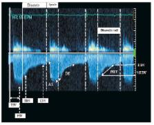
          Twenty-three patients, including 16 males and seven females with a mean age of 26.14 ± 10.17 years, were enrolled in this study. Except for the mean velocity and mean pressure gradient of the abdominal aorta, the values of the other indices of the abdominal/descending aorta showed enough change after stenting to indicate significant diagnostic accuracy for detecting aortic coarctation. The velocity–time integral and the pressure half-time were among the indices with the highest accuracy rates for this purpose (all p < 0.001).

          Conclusion

          Post-stenting echocardiographic profiles could provide a reliable reference value of the normal aortic haemodynamics as a unique identification of each patient and it is presumed that these indices could be used as reliable indicators of response to treatment.

          Related collections

          Most cited references40

          • Record: found
          • Abstract: not found
          • Article: not found

          Arterial assessment by Doppler-shift ultrasound.

            Bookmark
            • Record: found
            • Abstract: found
            • Article: not found

            Endovascular stents for coarctation of the aorta: initial results and intermediate-term follow-up.

            The aim of this study was to evaluate the use of endovascular stents in native and recurrent coarctation of the aorta (CoA). Stents have been used successfully in various locations. Their use in CoA can be an alternative to surgery or balloon angioplasty (BA). Thirty-four patients with CoA (13 native and 21 re-coarctation after surgery or BA) with a mean age of 16 +/- 8 years (range 4 to 36 years) underwent attempted stent implantation between 1993 and 1999. Successful outcome was defined as peak systolic pressure gradient after stent implantation < 20 mm Hg. Stents were implanted in 33/34 patients, and successful outcome occurred in 32/33 patients. Peak systolic pressure gradient decreased from 32 +/- 12 mm Hg to 4 +/- 11 mm Hg (p < 0.001). Coarctation site to descending aorta diameter ratio increased from 0.46 +/- 0.16 to 0.92 +/- 0.16 (p < 0.001). Two patients underwent successful stent re-dilation 16 and 21 months after initial implantation. Six patients (18%) developed complications, including two patients who underwent surgery. Follow-up for 29 +/- 17 months (range: 5 to 81 months) demonstrated no evidence of re-coarctation, aneurysm formation, stent displacement or fracture. Systolic blood pressure (SBP) decreased from 136 +/- 21 mm Hg before stent placement to 122 +/- 19 mm Hg at follow-up (p = 0.002). The SBP gradient decreased from 39 +/- 18 mm Hg to 4 +/- 6 mm Hg, and peak Doppler gradient decreased from 51 +/- 26 mm Hg to 13 +/- 11 mm Hg at follow-up (p < 0.001). Intravascular stent placement for native and recurrent CoA has excellent results in the short and intermediate terms. Long-term outcome remains to be evaluated.
              Bookmark
              • Record: found
              • Abstract: found
              • Article: not found

              Implantation and intermediate-term follow-up of stents in congenital heart disease.

              Balloon-expandable stents (Johnson and Johnson Interventional Systems) have been in use for congenital heart disease since late 1989. They have made possible treatment in previously untreatable branch pulmonary artery stenoses and systemic venous stenosis. The purpose of this report is to detail the results and intermediate-term follow-up of stents used for treatment of congenital heart disease. Eighty-five patients underwent placement of 121 stents in Houston and Boston. Fifty-eight patients had stents put in pulmonary arteries, nine had stents in conduits or outflow tracts, and 21 had stents in venous stenoses or narrowed Fontan anastomoses. (Three patients had stents in two locations.) These stent procedures resulted in gradient reduction from 55.2 +/- 33.3 to 14.2 +/- 13.5 mm Hg in pulmonary arteries, from 41.4 +/- 26.0 to 20.7 +/- 17.0 mm Hg in conduits or outflow tracts, and from 9.8 +/- 6.9 to 2.4 +/- 3.1 mm Hg in venous stenoses or Fontan anastomoses. Diameter of narrowings increased from 4.6 +/- 2.3 to 11.3 +/- 3.2 mm in the pulmonary artery, from 8.8 +/- 3.6 to 12.7 +/- 2.6 in conduits, and from 3.8 +/- 2.9 to 11.3 +/- 2.8 in venous stenoses. Follow-up has shown stent fracture in one patient, restenosis in one, and sudden death in one. Recatheterization has been done in 38 patients an average of 8.6 months after stent installation. Compared with immediately postimplant data, there was no significant change in luminal diameter or pressure gradient. Redilation was performed in 14 patients (17 stents) 1 week to 24 months after implantation (mean, 10.2 months), with a small but significant increase in stenosis diameter. We conclude that stent treatment of vascular stenoses in congenital heart disease retains efficacy at medium-term follow-up and offers a much-improved outlook for patients with these lesions.
                Bookmark

                Author and article information

                Contributors
                Journal
                Cardiovasc J Afr
                Cardiovasc J Afr
                TBC
                Cardiovascular Journal of Africa
                Clinics Cardive Publishing
                1995-1892
                1680-0745
                October 2012
                : 23
                : 9
                : 483-490
                Affiliations
                Department of Cardiology, Rasoul-e-Akram Hospital, Tehran
                University of Medical Sciences (TUMS), Tehran, Iran
                Firoozgar Clinical Research Development Center (FCRDC), Firoozgar Hospital, Tehran University of Medical Sciences (TUMS), Tehran, Iran
                Department of Echocardiography and Cardiology, Shaheed Rajaie Cardiovascular Medical and Research Centre, Tehran
                University of Medical Sciences (TUMS), Tehran, Iran
                Department of Echocardiography and Cardiology, Shaheed Rajaie Cardiovascular Medical and Research Centre, Tehran
                University of Medical Sciences (TUMS), Tehran, Iran
                Department of Surgery, Firoozgar Hospital, Tehran
                University of Medical Sciences (TUMS), Tehran, Iran
                Medical Students’ Cardiology Research Centre, Shaheed
                Rajaie Cardiovascular Medical and Research Centre, Tehran
                University of Medical Sciences (TUMS), Tehran, Iran
                Medical Students’ Research Committee (MSRC), Tehran
                University of Medical Sciences (TUMS), and Rasoul-e-Akram Hospital, Tehran, Iran
                Article
                10.5830/CVJA-2012-044
                3721869
                23108515
                ecd1562a-0537-4a41-b4e5-51ec919e193b
                Copyright © 2010 Clinics Cardive Publishing

                This is an open-access article distributed under the terms of the Creative Commons Attribution License, which permits unrestricted use, distribution, and reproduction in any medium, provided the original work is properly cited.

                History
                : 2 October 2011
                : 11 May 2012
                Categories
                Cardiovascular Topics

                aortic coarctation,doppler echocardiography,index,diagnostic values

                Comments

                Comment on this article