6
views
0
recommends
+1 Recommend
0 collections
    0
    shares
      • Record: found
      • Abstract: found
      • Article: found
      Is Open Access

      Anatomical basis and physiological role of cerebrospinal fluid transport through the murine cribriform plate

      research-article

      Read this article at

      Bookmark
          There is no author summary for this article yet. Authors can add summaries to their articles on ScienceOpen to make them more accessible to a non-specialist audience.

          Abstract

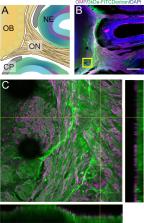
          Cerebrospinal fluid (CSF) flows through the brain, transporting chemical signals and removing waste. CSF production in the brain is balanced by a constant outflow of CSF, the anatomical basis of which is poorly understood. Here, we characterized the anatomy and physiological function of the CSF outflow pathway along the olfactory sensory nerves through the cribriform plate, and into the nasal epithelia. Chemical ablation of olfactory sensory nerves greatly reduced outflow of CSF through the cribriform plate. The reduction in CSF outflow did not cause an increase in intracranial pressure (ICP), consistent with an alteration in the pattern of CSF drainage or production. Our results suggest that damage to olfactory sensory neurons (such as from air pollution) could contribute to altered CSF turnover and flow, providing a potential mechanism for neurological diseases.

          Related collections

          Most cited references105

          • Record: found
          • Abstract: found
          • Article: not found

          Intranasal delivery of biologics to the central nervous system.

          Treatment of central nervous system (CNS) diseases is very difficult due to the blood-brain barrier's (BBB) ability to severely restrict entry of all but small, non-polar compounds. Intranasal administration is a non-invasive method of drug delivery which may bypass the BBB to allow therapeutic substances direct access to the CNS. Intranasal delivery of large molecular weight biologics such as proteins, gene vectors, and stem cells is a potentially useful strategy to treat a variety of diseases/disorders of the CNS including stroke, Parkinson's disease, multiple sclerosis, Alzheimer's disease, epilepsy, and psychiatric disorders. Here we give an overview of relevant nasal anatomy and physiology and discuss the pathways and mechanisms likely involved in drug transport from the nasal epithelium to the CNS. Finally we review both pre-clinical and clinical studies involving intranasal delivery of biologics to the CNS. Copyright © 2011 Elsevier B.V. All rights reserved.
            Bookmark
            • Record: found
            • Abstract: found
            • Article: not found

            SeeDB: a simple and morphology-preserving optical clearing agent for neuronal circuit reconstruction.

            We report a water-based optical clearing agent, SeeDB, which clears fixed brain samples in a few days without quenching many types of fluorescent dyes, including fluorescent proteins and lipophilic neuronal tracers. Our method maintained a constant sample volume during the clearing procedure, an important factor for keeping cellular morphology intact, and facilitated the quantitative reconstruction of neuronal circuits. Combined with two-photon microscopy and an optimized objective lens, we were able to image the mouse brain from the dorsal to the ventral side. We used SeeDB to describe the near-complete wiring diagram of sister mitral cells associated with a common glomerulus in the mouse olfactory bulb. We found the diversity of dendrite wiring patterns among sister mitral cells, and our results provide an anatomical basis for non-redundant odor coding by these neurons. Our simple and efficient method is useful for imaging intact morphological architecture at large scales in both the adult and developing brains.
              Bookmark
              • Record: found
              • Abstract: found
              • Article: not found

              Correlations of neuronal and microvascular densities in murine cortex revealed by direct counting and colocalization of nuclei and vessels.

              It is well known that the density of neurons varies within the adult brain. In neocortex, this includes variations in neuronal density between different lamina as well as between different regions. Yet the concomitant variation of the microvessels is largely uncharted. Here, we present automated histological, imaging, and analysis tools to simultaneously map the locations of all neuronal and non-neuronal nuclei and the centerlines and diameters of all blood vessels within thick slabs of neocortex from mice. Based on total inventory measurements of different cortical regions ( approximately 10(7) cells vectorized across brains), these methods revealed: (1) In three dimensions, the mean distance of the center of neuronal somata to the closest microvessel was 15 mum. (2) Volume samples within lamina of a given region show that the density of microvessels does not match the strong laminar variation in neuronal density. This holds for both agranular and granular cortex. (3) Volume samples in successive radii from the midline to the ventral-lateral edge, where each volume summed the number of cells and microvessels from the pia to the white matter, show a significant correlation between neuronal and microvessel densities. These data show that while neuronal and vascular densities do not track each other on the 100 mum scale of cortical lamina, they do track each other on the 1-10 mm scale of the cortical mantle. The absence of a disproportionate density of blood vessels in granular lamina is argued to be consistent with the initial locus of functional brain imaging signals.
                Bookmark

                Author and article information

                Contributors
                Role: Reviewing Editor
                Role: Senior Editor
                Journal
                eLife
                Elife
                eLife
                eLife
                eLife Sciences Publications, Ltd
                2050-084X
                07 May 2019
                2019
                : 8
                : e44278
                Affiliations
                [1 ]deptCellular and Developmental Biology Graduate Program Pennsylvania State University University ParkUnited States
                [2 ]deptDepartment of Engineering Science and Mechanics Pennsylvania State University University ParkUnited States
                [3 ]deptDepartment of Physics Pennsylvania State University University ParkUnited States
                [4 ]deptDepartment of Biomedical Engineering Pennsylvania State University University ParkUnited States
                [5 ]deptDepartment of Anthropology Pennsylvania State University University ParkUnited States
                [6 ]deptDepartment of Neurosurgery Pennsylvania State University University ParkUnited States
                Emory University United States
                Emory University United States
                Emory University United States
                University of Southampton United Kingdom
                University of Wisconsin-Madison United States
                OHSU United States
                Author information
                http://orcid.org/0000-0001-8093-5938
                http://orcid.org/0000-0003-4500-813X
                https://orcid.org/0000-0002-7483-7378
                Article
                44278
                10.7554/eLife.44278
                6524970
                31063132
                ec91be1e-2f0d-417d-9e8a-8d3cfbf50cff
                © 2019, Norwood et al

                This article is distributed under the terms of the Creative Commons Attribution License, which permits unrestricted use and redistribution provided that the original author and source are credited.

                History
                : 10 December 2018
                : 06 May 2019
                Funding
                Funded by: FundRef http://dx.doi.org/10.13039/100000002, National Institutes of Health;
                Award ID: F31NS105461
                Award Recipient :
                Funded by: FundRef http://dx.doi.org/10.13039/100008321, Pennsylvania State University;
                Award ID: Huck Graduate Enrichment Grant
                Award Recipient :
                Funded by: FundRef http://dx.doi.org/10.13039/100000001, National Science Foundation;
                Award ID: CBET1705854
                Award Recipient :
                Funded by: FundRef http://dx.doi.org/10.13039/100000872, McKnight Endowment Fund for Neuroscience;
                Award Recipient :
                Funded by: FundRef http://dx.doi.org/10.13039/100000002, National Institutes of Health;
                Award ID: R01NS078168
                Award Recipient :
                Funded by: FundRef http://dx.doi.org/10.13039/100000002, National Institutes of Health;
                Award ID: P01HD078233
                Award Recipient :
                The funders had no role in study design, data collection and interpretation, or the decision to submit the work for publication.
                Categories
                Research Article
                Neuroscience
                Custom metadata
                Chemical ablation of olfactory sensory neurons blocks cerebrospinal fluid outflow into the nasal epithelium, leading to alterations of cerebrospinal fluid dynamics.

                Life sciences
                cribriform,olfatory nerve,cerebrospinal fluid,intracranial pressure,anosmia,mouse
                Life sciences
                cribriform, olfatory nerve, cerebrospinal fluid, intracranial pressure, anosmia, mouse

                Comments

                Comment on this article