4
views
0
recommends
+1 Recommend
0 collections
    0
    shares
      • Record: found
      • Abstract: found
      • Article: found
      Is Open Access

      Validation of clinical acceptability of deep-learning-based automated segmentation of organs-at-risk for head-and-neck radiotherapy treatment planning

      research-article

      Read this article at

      Bookmark
          There is no author summary for this article yet. Authors can add summaries to their articles on ScienceOpen to make them more accessible to a non-specialist audience.

          Abstract

          Introduction

          Organ-at-risk segmentation for head and neck cancer radiation therapy is a complex and time-consuming process (requiring up to 42 individual structure, and may delay start of treatment or even limit access to function-preserving care. Feasibility of using a deep learning (DL) based autosegmentation model to reduce contouring time without compromising contour accuracy is assessed through a blinded randomized trial of radiation oncologists (ROs) using retrospective, de-identified patient data.

          Methods

          Two head and neck expert ROs used dedicated time to create gold standard (GS) contours on computed tomography (CT) images. 445 CTs were used to train a custom 3D U-Net DL model covering 42 organs-at-risk, with an additional 20 CTs were held out for the randomized trial. For each held-out patient dataset, one of the eight participant ROs was randomly allocated to review and revise the contours produced by the DL model, while another reviewed contours produced by a medical dosimetry assistant (MDA), both blinded to their origin. Time required for MDAs and ROs to contour was recorded, and the unrevised DL contours, as well as the RO-revised contours by the MDAs and DL model were compared to the GS for that patient.

          Results

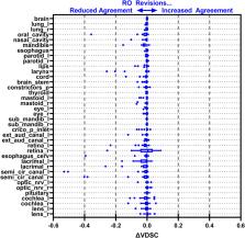
          Mean time for initial MDA contouring was 2.3 hours (range 1.6-3.8 hours) and RO-revision took 1.1 hours (range, 0.4-4.4 hours), compared to 0.7 hours (range 0.1-2.0 hours) for the RO-revisions to DL contours. Total time reduced by 76% (95%-Confidence Interval: 65%-88%) and RO-revision time reduced by 35% (95%-CI,-39%-91%). All geometric and dosimetric metrics computed, agreement with GS was equivalent or significantly greater (p<0.05) for RO-revised DL contours compared to the RO-revised MDA contours, including volumetric Dice similarity coefficient (VDSC), surface DSC, added path length, and the 95%-Hausdorff distance. 32 OARs (76%) had mean VDSC greater than 0.8 for the RO-revised DL contours, compared to 20 (48%) for RO-revised MDA contours, and 34 (81%) for the unrevised DL OARs.

          Conclusion

          DL autosegmentation demonstrated significant time-savings for organ-at-risk contouring while improving agreement with the institutional GS, indicating comparable accuracy of DL model. Integration into the clinical practice with a prospective evaluation is currently underway.

          Related collections

          Most cited references69

          • Record: found
          • Abstract: found
          • Article: not found

          Global cancer statistics 2020: GLOBOCAN estimates of incidence and mortality worldwide for 36 cancers in 185 countries

          This article provides an update on the global cancer burden using the GLOBOCAN 2020 estimates of cancer incidence and mortality produced by the International Agency for Research on Cancer. Worldwide, an estimated 19.3 million new cancer cases (18.1 million excluding nonmelanoma skin cancer) and almost 10.0 million cancer deaths (9.9 million excluding nonmelanoma skin cancer) occurred in 2020. Female breast cancer has surpassed lung cancer as the most commonly diagnosed cancer, with an estimated 2.3 million new cases (11.7%), followed by lung (11.4%), colorectal (10.0 %), prostate (7.3%), and stomach (5.6%) cancers. Lung cancer remained the leading cause of cancer death, with an estimated 1.8 million deaths (18%), followed by colorectal (9.4%), liver (8.3%), stomach (7.7%), and female breast (6.9%) cancers. Overall incidence was from 2-fold to 3-fold higher in transitioned versus transitioning countries for both sexes, whereas mortality varied <2-fold for men and little for women. Death rates for female breast and cervical cancers, however, were considerably higher in transitioning versus transitioned countries (15.0 vs 12.8 per 100,000 and 12.4 vs 5.2 per 100,000, respectively). The global cancer burden is expected to be 28.4 million cases in 2040, a 47% rise from 2020, with a larger increase in transitioning (64% to 95%) versus transitioned (32% to 56%) countries due to demographic changes, although this may be further exacerbated by increasing risk factors associated with globalization and a growing economy. Efforts to build a sustainable infrastructure for the dissemination of cancer prevention measures and provision of cancer care in transitioning countries is critical for global cancer control.
            Bookmark
            • Record: found
            • Abstract: found
            • Article: found
            Is Open Access

            Metrics for evaluating 3D medical image segmentation: analysis, selection, and tool

            Background Medical Image segmentation is an important image processing step. Comparing images to evaluate the quality of segmentation is an essential part of measuring progress in this research area. Some of the challenges in evaluating medical segmentation are: metric selection, the use in the literature of multiple definitions for certain metrics, inefficiency of the metric calculation implementations leading to difficulties with large volumes, and lack of support for fuzzy segmentation by existing metrics. Result First we present an overview of 20 evaluation metrics selected based on a comprehensive literature review. For fuzzy segmentation, which shows the level of membership of each voxel to multiple classes, fuzzy definitions of all metrics are provided. We present a discussion about metric properties to provide a guide for selecting evaluation metrics. Finally, we propose an efficient evaluation tool implementing the 20 selected metrics. The tool is optimized to perform efficiently in terms of speed and required memory, also if the image size is extremely large as in the case of whole body MRI or CT volume segmentation. An implementation of this tool is available as an open source project. Conclusion We propose an efficient evaluation tool for 3D medical image segmentation using 20 evaluation metrics and provide guidelines for selecting a subset of these metrics that is suitable for the data and the segmentation task.
              Bookmark
              • Record: found
              • Abstract: not found
              • Article: not found

              Nasa-Task Load Index (NASA-TLX); 20 Years Later

              S. G. Hart (2006)
                Bookmark

                Author and article information

                Contributors
                Journal
                Front Oncol
                Front Oncol
                Front. Oncol.
                Frontiers in Oncology
                Frontiers Media S.A.
                2234-943X
                06 April 2023
                2023
                : 13
                : 1137803
                Affiliations
                [1] 1 Department of Radiation Oncology, Mayo Clinic , Rochester, MN, United States
                [2] 2 Department of Health Sciences Research, Mayo Clinic , Phoenix, AZ, United States
                [3] 3 Department of Radiation Oncology, Mayo Clinic , Phoenix, AZ, United States
                [4] 4 Department of Radiation Oncology, Mayo Clinic , Jacksonville, FL, United States
                [5] 5 Google Health , Mountain View, CA, United States
                [6] 6 Research Services, Comprehensive Cancer Center, Mayo Clinic , Rochester, MN, United States
                [7] 7 Robert D. and Patricia E. Kern Center for the Science of Health Care Delivery, Mayo Clinic , Rochester, MN, United States
                Author notes

                Edited by: Giuseppe Carlo Iorio, University of Turin, Italy

                Reviewed by: Roberta Grassi, University of Campania Luigi Vanvitelli, Italy; Donatella Caivano, Sant’Andrea University Hospital, Sapienza University of Rome, Italy; Matthew Field, University of New South Wales, Australia

                *Correspondence: J. John Lucido, lucido.joseph@ 123456mayo.edu

                This article was submitted to Radiation Oncology, a section of the journal Frontiers in Oncology

                Article
                10.3389/fonc.2023.1137803
                10115982
                37091160
                ebf1cc8b-3161-4d55-8a57-84e3944c1ca5
                Copyright © 2023 Lucido, DeWees, Leavitt, Anand, Beltran, Brooke, Buroker, Foote, Foss, Gleason, Hodge, Hughes, Hunzeker, Laack, Lenz, Livne, Morigami, Moseley, Undahl, Patel, Tryggestad, Walker, Zverovitch and Patel

                This is an open-access article distributed under the terms of the Creative Commons Attribution License (CC BY). The use, distribution or reproduction in other forums is permitted, provided the original author(s) and the copyright owner(s) are credited and that the original publication in this journal is cited, in accordance with accepted academic practice. No use, distribution or reproduction is permitted which does not comply with these terms.

                History
                : 04 January 2023
                : 24 March 2023
                Page count
                Figures: 9, Tables: 4, Equations: 0, References: 69, Pages: 15, Words: 6890
                Categories
                Oncology
                Original Research

                Oncology & Radiotherapy
                deep learning,autosegmentation,head and neck cancer,radiation therapy,clinical validation,comprehensive,organs-at-risk

                Comments

                Comment on this article