0
views
0
recommends
+1 Recommend
0 collections
    0
    shares
      • Record: found
      • Abstract: found
      • Article: found
      Is Open Access

      A Unique Upside-down Flip-lid Type of Lateral Tibial Plateau Fracture Fragment in a Bicondylar Tibial Plateau Fracture Managed with Dual Plates and Raft Screws

      case-report

      Read this article at

      Bookmark
          There is no author summary for this article yet. Authors can add summaries to their articles on ScienceOpen to make them more accessible to a non-specialist audience.

          Abstract

          Introduction:

          Bicondylar fractures are relatively common, yet those involving an elevated lateral tibial condyle fragment pose a unique challenge due to their atypical presentation. Existing classification systems inadequately describe this elevation, leading to varied terminology like “flip lid” or “reverse-Schatzker type” fractures in the literature.

          Case Report:

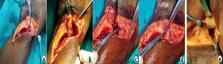
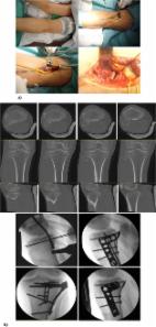
          We present a case where the anterolateral osteochondral fragment was elevated and inverted, resulting from a rare mechanism where the left knee was crushed between two vehicles without axial force. This unusual mechanism spared typical signs of condylar widening or depression, with the fragment elevating but remaining submeniscal. This presented a challenge for fixation. The rotated fragment was accessed through an anterolateral approach with submeniscal arthrotomy, reduced, and fixed using raft screws of the lateral locking plate. The medial plateau fracture was stabilized through a posteromedial approach using an antiglide plate. Follow-up at 14 months showed satisfactory outcomes.

          Conclusion:

          Unique injury mechanisms can give rise to distinct fracture types. When X-rays depict an elevated rather than depressed articular surface, suspicion should arise for an elevated, “popped up,” or flip lid type fragment. Such cases require a high index of suspicion and a thorough preoperative evaluation using both X-rays and CT scans. Submeniscal arthrotomy is essential to assess meniscus integrity and allow direct visualization of the fracture fragment. Successful outcomes in managing these fractures are based on accurate diagnosis, thorough preoperative planning, and adherence to internal fixation principles.

          Related collections

          Most cited references21

          • Record: found
          • Abstract: found
          • Article: not found

          Three-column fixation for complex tibial plateau fractures.

          1) To introduce a computed tomography-based "three-column fixation" concept; and 2) to evaluate clinical outcomes (by using a column-specific fixation technique) for complex tibial plateau fractures (Schatzker classification Types V and VI). Prospective cohort study. Level 1 trauma center. Twenty-nine cases of complex tibial plateau fractures were included. Based on routine x-ray and computed tomography images, all the fractures were classified as a "three-column fracture," which means at least one separate fragment was found in lateral, medial, and posterior columns in the proximal tibia (Schatzker classification Types V and VI). The patients were operated on in a "floating position" with a combined approach, an inverted L-shaped posterior approach combined with an anterior-lateral approach. All three columns of fractures were fixed. Operative time, blood loss, quality of reduction and alignment, fracture healing, complications, and functional outcomes based on Hospital for Special Surgery score and lower-extremity measure were recorded. All the cases were followed for average 27.3 months (range, 24-36 months). All the cases had satisfactory reduction except one case, which had a 4-mm stepoff at the anterior ridge of the tibial plateau postoperatively. No case of secondary articular depression was found. One case had secondary varus deformity, one case had secondary valgus deformity, and two cases of screw loosening occurred postoperatively. No revision surgery was performed. Two cases had culture-negative wound drainage. No infection was noted. The average radiographic bony union time and full weightbearing time were 13.1 weeks (range, 11-16 weeks) and 16.7 weeks (range, 12-24 weeks), respectively. The mean Short Form 36, Hospital for Special Surgery score, and lower-extremity measure at 24 months postoperatively were 89 (range, 80-98), 90 (range, 84-98), and 87 (range, 80-95), respectively. The average range of motion of the affected knee was 2.7° to 123.4° at 2 years after the operation. Three-column fixation is a new fixation concept in treating complex tibial plateau fractures, which is especially useful for multiplanar fractures involving the posterior column. The combination of posterior and anterior-lateral approaches is a safe and effective way to have direct reduction and satisfactory fixation for such difficult tibial plateau fractures.
            Bookmark
            • Record: found
            • Abstract: found
            • Article: found
            Is Open Access

            Treatment strategy for tibial plateau fractures: an update

            Tibial plateau fractures are complex injuries produced by high- or low-energy trauma. They principally affect young adults or the ‘third age’ population. These fractures usually have associated soft-tissue lesions that will affect their treatment. Sequential (staged) treatment (external fixation followed by definitive osteosynthesis) is recommended in more complex fracture patterns. But one should remember that any type of tibial plateau fracture can present with soft-tissue complications. Typically the Schatzker or AO/OTA classification is used, but the concept of the proximal tibia as a three-column structure and the detailed study of the posteromedial and posterolateral fragment morphology has changed its treatment strategy. Limb alignment and articular surface restoration, allowing early knee motion, are the main goals of surgical treatment. Partially articular factures can be treated by minimally-invasive methods and arthroscopy is useful to assist and control the fracture reduction and to treat intra-articular soft-tissue injuries. Open reduction and internal fixation (ORIF) is the gold standard treatment for these fractures. Complex articular fractures can be treated by ring external fixators and minimally-invasive osteosynthesis (EFMO) or by ORIF. EFMO can be related to suboptimal articular reduction; however, outcome analysis shows results that are equal to, or even superior to, ORIF. The ORIF strategy should also include the optimal reduction of the articular surface. Anterolateral and anteromedial surgical approaches do not permit adequate reduction and fixation of posterolateral and posteromedial fragments. To achieve this, it is necessary to reduce and fix them through specific posterolateral or posteromedial approaches that allow optimal reduction and plate/screw placement. Some authors have also suggested that primary total knee arthroplasty could be an option in specific patients and with specific fracture patterns. Cite this article: Prat-Fabregat S, Camacho-Carrasco P. Treatment strategy for tibial plateau fractures: an update. EFORT Open Rev 2016;1:225-232. DOI: 10.1302/2058-5241.1.000031.
              Bookmark
              • Record: found
              • Abstract: found
              • Article: not found

              Frequency and fracture morphology of the posteromedial fragment in bicondylar tibial plateau fracture patterns.

              Bicondylar tibial plateau fracture management remains therapeutically challenging, partly because of multiplanar articular comminution. This study was performed to evaluate the frequency and morphologic characteristics of the posteromedial fragment in this injury pattern. Retrospective chart and radiographic review. Urban Level 1 university trauma center. Fifty-seven patients sustaining 57 Orthopedic Trauma Association (OTA) C-Type bicondylar tibial plateau fractures formed the study group. Between May 2000 and March 2003, 170 OTA C-Type bicondylar tibial plateau fractures were identified using an orthopaedic database. One hundred and forty-six fractures had computed tomographic (CT) scans performed prior to definitive fixation and were reviewed using the Picture Archiving and Communication System (PACS). Sixty-six (45.2%) injuries had fractures that involved the medial articular surface. Nine with suboptimal CTs were excluded, leaving 57 injuries for review. Forty-two patients demonstrated coronal plane posteromedial fragments. Morphologic evaluation of the posteromedial fragment included articular surface area, maximum posterior cortical height (PCH), and sagittal fracture angle (SFA). Forty-two of 57 injuries (74%) demonstrated a posteromedial fragment that comprised a mean of 58% of the articular surface of the medial tibial plateau (range, 19%-98%) and a mean of 23% of the entire tibial plateau articular surface (range, 8%-47%). Mean posteromedial fragment height was 42 mm (range, 16-59 mm), and mean sagittal fracture angle was 81 degrees (range, 33 degrees to 112 degrees). Six patients demonstrated fracture patterns not accurately identified by the AO/OTA (Arbeitsgemeinschaft für Osteosynthesefragen/Orthopaedic Trauma Association) fracture classification system. A posteromedial fragment was observed in nearly one third of the bicondylar plateau fractures evaluated. The morphologic features of this fragment may have clinical implications when using currently available laterally applied fixed-angle screw/plate implants to stabilize these injuries. Alternate or supplementary fixation methods may be required when managing this injury pattern.
                Bookmark

                Author and article information

                Journal
                J Orthop Case Rep
                J Orthop Case Rep
                Journal of Orthopaedic Case Reports
                Indian Orthopaedic Research Group (India )
                2250-0685
                2321-3817
                August 2024
                August 2024
                : 14
                : 8
                : 25-29
                Affiliations
                [1 ]Department of Orthopaedics, Paras HMRI Hospital, Patna, Bihar, India
                Author notes
                Address of Correspondence: Dr. Janki Sharan Bhadani, Department of Orthopaedics, Paras HMRI Hospital, Patna, Bihar, India. E-mail: jsbhadani@ 123456gmail.com
                Article
                JOCR-14-25
                10.13107/jocr.2024.v14.i08.4634
                11327674
                39157483
                ebc7750a-fa70-4754-bd61-a56592d3fd50
                Copyright: © Indian Orthopaedic Research Group

                This is an open access journal, and articles are distributed under the terms of the Creative Commons Attribution-NonCommercial-ShareAlike 4.0 Unported, which allows others to remix, tweak, and build upon the work non-commercially, as long as appropriate credit is given and the new creations are licensed under the identical terms

                History
                : 06 April 2024
                : 22 May 2024
                : June 2024
                Categories
                Case Report

                elevated,popped up,flip lid,reverse tibial plateau fractures

                Comments

                Comment on this article