19
views
0
recommends
+1 Recommend
0 collections
    0
    shares
      • Record: found
      • Abstract: found
      • Article: found
      Is Open Access

      The Role of the Antioxidant Response in Mitochondrial Dysfunction in Degenerative Diseases: Cross-Talk between Antioxidant Defense, Autophagy, and Apoptosis

      review-article

      Read this article at

      Bookmark
          There is no author summary for this article yet. Authors can add summaries to their articles on ScienceOpen to make them more accessible to a non-specialist audience.

          Abstract

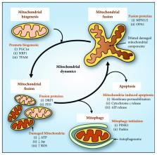
          The mitochondrion is an essential organelle important for the generation of ATP for cellular function. This is especially critical for cells with high energy demands, such as neurons for signal transmission and cardiomyocytes for the continuous mechanical work of the heart. However, deleterious reactive oxygen species are generated as a result of mitochondrial electron transport, requiring a rigorous activation of antioxidative defense in order to maintain homeostatic mitochondrial function. Indeed, recent studies have demonstrated that the dysregulation of antioxidant response leads to mitochondrial dysfunction in human degenerative diseases affecting the nervous system and the heart. In this review, we outline and discuss the mitochondrial and oxidative stress factors causing degenerative diseases, such as Alzheimer's disease, Parkinson's disease, amyotrophic lateral sclerosis, Huntington's disease, and Friedreich's ataxia. In particular, the pathological involvement of mitochondrial dysfunction in relation to oxidative stress, energy metabolism, mitochondrial dynamics, and cell death will be explored. Understanding the pathology and the development of these diseases has highlighted novel regulators in the homeostatic maintenance of mitochondria. Importantly, this offers potential therapeutic targets in the development of future treatments for these degenerative diseases.

          Related collections

          Most cited references204

          • Record: found
          • Abstract: found
          • Article: not found

          Direct activation of Bax by p53 mediates mitochondrial membrane permeabilization and apoptosis.

          The tumor suppressor p53 exerts its anti-neoplastic activity primarily through the induction of apoptosis. We found that cytosolic localization of endogenous wild-type or trans-activation-deficient p53 was necessary and sufficient for apoptosis. p53 directly activated the proapoptotic Bcl-2 protein Bax in the absence of other proteins to permeabilize mitochondria and engage the apoptotic program. p53 also released both proapoptotic multidomain proteins and BH3-only proteins [Proapoptotic Bcl-2 family proteins that share only the third Bcl-2 homology domain (BH3)] that were sequestered by Bcl-xL. The transcription-independent activation of Bax by p53 occurred with similar kinetics and concentrations to those produced by activated Bid. We propose that when p53 accumulates in the cytosol, it can function analogously to the BH3-only subset of proapoptotic Bcl-2 proteins to activate Bax and trigger apoptosis.
            Bookmark
            • Record: found
            • Abstract: found
            • Article: not found

            Nix is a selective autophagy receptor for mitochondrial clearance.

            Autophagy is the cellular homeostatic pathway that delivers large cytosolic materials for degradation in the lysosome. Recent evidence indicates that autophagy mediates selective removal of protein aggregates, organelles and microbes in cells. Yet, the specificity in targeting a particular substrate to the autophagy pathway remains poorly understood. Here, we show that the mitochondrial protein Nix is a selective autophagy receptor by binding to LC3/GABARAP proteins, ubiquitin-like modifiers that are required for the growth of autophagosomal membranes. In cultured cells, Nix recruits GABARAP-L1 to damaged mitochondria through its amino-terminal LC3-interacting region. Furthermore, ablation of the Nix:LC3/GABARAP interaction retards mitochondrial clearance in maturing murine reticulocytes. Thus, Nix functions as an autophagy receptor, which mediates mitochondrial clearance after mitochondrial damage and during erythrocyte differentiation.
              Bookmark
              • Record: found
              • Abstract: found
              • Article: found
              Is Open Access

              PINK1 is activated by mitochondrial membrane potential depolarization and stimulates Parkin E3 ligase activity by phosphorylating Serine 65

              Summary Missense mutations in PTEN-induced kinase 1 (PINK1) cause autosomal-recessive inherited Parkinson's disease (PD). We have exploited our recent discovery that recombinant insect PINK1 is catalytically active to test whether PINK1 directly phosphorylates 15 proteins encoded by PD-associated genes as well as proteins reported to bind PINK1. We have discovered that insect PINK1 efficiently phosphorylates only one of these proteins, namely the E3 ligase Parkin. We have mapped the phosphorylation site to a highly conserved residue within the Ubl domain of Parkin at Ser65. We show that human PINK1 is specifically activated by mitochondrial membrane potential (Δψm) depolarization, enabling it to phosphorylate Parkin at Ser65. We further show that phosphorylation of Parkin at Ser65 leads to marked activation of its E3 ligase activity that is prevented by mutation of Ser65 or inactivation of PINK1. We provide evidence that once activated, PINK1 autophosphorylates at several residues, including Thr257, which is accompanied by an electrophoretic mobility band-shift. These results provide the first evidence that PINK1 is activated following Δψm depolarization and suggest that PINK1 directly phosphorylates and activates Parkin. Our findings indicate that monitoring phosphorylation of Parkin at Ser65 and/or PINK1 at Thr257 represent the first biomarkers for examining activity of the PINK1-Parkin signalling pathway in vivo. Our findings also suggest that small molecule activators of Parkin that mimic the effect of PINK1 phosphorylation may confer therapeutic benefit for PD.
                Bookmark

                Author and article information

                Contributors
                Journal
                Oxid Med Cell Longev
                Oxid Med Cell Longev
                OMCL
                Oxidative Medicine and Cellular Longevity
                Hindawi
                1942-0900
                1942-0994
                2019
                7 April 2019
                : 2019
                : 6392763
                Affiliations
                Molecular Pharmacology and Pathology Program, Department of Pathology and Bosch Institute, Medical Foundation Building (K25), University of Sydney, Sydney, New South Wales 2006, Australia
                Author notes

                Guest Editor: Livia Hool

                Author information
                http://orcid.org/0000-0003-0693-7833
                http://orcid.org/0000-0002-5043-9586
                http://orcid.org/0000-0003-0960-6415
                Article
                10.1155/2019/6392763
                6476015
                31057691
                ebc37cc1-f5b6-405c-b907-8997203c4698
                Copyright © 2019 Michael L.-H. Huang et al.

                This is an open access article distributed under the Creative Commons Attribution License, which permits unrestricted use, distribution, and reproduction in any medium, provided the original work is properly cited.

                History
                : 13 November 2018
                : 18 January 2019
                : 11 February 2019
                Funding
                Funded by: National Health and Medical Research Council
                Award ID: APP1074033
                Award ID: APP1062607
                Award ID: APP1128152
                Award ID: APP1048972
                Funded by: University of Sydney
                Funded by: Muscular Dystrophy Association
                Award ID: MDA417129
                Funded by: Cancer Australia
                Funded by: Cure Cancer Australia Foundation
                Categories
                Review Article

                Molecular medicine
                Molecular medicine

                Comments

                Comment on this article