25
views
0
recommends
+1 Recommend
0 collections
    0
    shares
      • Record: found
      • Abstract: found
      • Article: found
      Is Open Access

      Inverted ILM peeling for idiopathic and other etiology macular holes

      letter

      Read this article at

      Bookmark
          There is no author summary for this article yet. Authors can add summaries to their articles on ScienceOpen to make them more accessible to a non-specialist audience.

          Abstract

          Sir, We read the article titled “Surgical outcomes of inverted internal limiting membrane flap technique for large macular hole” by Prabhushanker Mahalingam et al.,[1] recently published in your esteem journal with great interest and would like to congratulate the authors for their work. We would like to contribute for the above said subject by sharing our experience of treating large/giant macular holes of various etiologies. We have performed vitrectomy, inverted flap internal limiting membrane (ILM) peeling and gas tamponade in 40 patients with the inclusion criteria being holes larger than 600 μm. Most patients had idiopathic macular hole (30 eyes - Fig. 1), others being vitreomacular traction syndrome (VMT) with associated macular hole (5 eyes), post cystoid macular edema (CME) (3 eyes) and post traumatic (2 eyes - Fig. 2) macular holes.
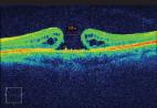
 Figure 1 OCT scan of a stage 4 idiopathic macular hole of minimal diameter of 970 microns. The inverted ILM flap is well seen in the immediate and late post operative period persisting upto months with gradual closure of the macular hole Figure 2 A large macular hole of size 1.3 mm secondary to blunt trauma, successfully treated with inverted ILM peeling with good visual recovery Of the 40 patients, 26 were phakic (23 had immature senile cataract, one had complicated cataract, one rosette cataract while the remaining one had a clear lens), and the remaining 14 were pseudophakic. Maximum pre-operative hole diameter was 1532 ± 168 μm. Fifteen of the 26 phakic eyes underwent combined cataract surgery, intraocular lens implantation along with macular hole surgery. Type 1 macular hole closure was achieved in 100% of the eyes at 1 month follow-up and the holes remained closed at 6 months. Optical coherence tomography showed persistent outer layer defects in the neuro-sensory retina in 5 eyes at the end of 1 month, of which 2 persisted even at 3 months and 6 months. The functional outcome in terms of visual improvement was noted in 25 eyes (62.5%). An increase of >4 lines was noted in 6 eyes, >2 lines in 13, <2 lines in 6 eyes while the remaining 15 maintained pre-operative vision. The mean pre-operative vision was 6/60 (1.00 LogMAR) which improved to 6/24 (0.602 LogMAR). Eyes with VMT associated macular hole improved the most with post-traumatic macular holes the least as compared to baseline. Visual improvement could be correlated with restoration of inner segment/outer segment (IS/OS) junction and this happened most in patients with VMT associated holes and idiopathic macular holes. Thus, we can conclude that inverted ILM flap technique is effective in closing large holes, as been shown both in our study as well as the pilot study by Michalewska Z et al.,[2] irrespective of the etiology. Type 1 closure can be achieved in a large number of patients associated with a modest visual improvement.

          Related collections

          Most cited references2

          • Record: found
          • Abstract: found
          • Article: not found

          Inverted internal limiting membrane flap technique for large macular holes.

          Large macular holes usually have an increased risk of surgical failure. Up to 44% of large macular holes remain open after 1 surgery. Another 19% to 39% of macular holes are flat-open after surgery. Flat-open macular holes are associated with limited visual acuity. This article presents a modification of the standard macular hole surgery to improve functional and anatomic outcomes in patients with large macular holes. A prospective, randomized clinical trial. Patients with macular holes larger than 400 μm were included. In group 1, 51 eyes of 40 patients underwent standard 3-port pars plana vitrectomy with air. In group 2, 50 eyes of 46 patients underwent a modification of the standard technique, called the inverted internal limiting membrane (ILM) flap technique. In the inverted ILM flap technique, instead of completely removing the ILM after trypan blue staining, a remnant attached to the margins of the macular hole was left in place. This ILM remnant was then inverted upside-down to cover the macular hole. Fluid-air exchange was then performed. Spectral optical coherence tomography and clinical examination were performed before surgery and postoperatively at 1 week and 1, 3, 6, and 12 months. Visual acuity and postoperative macular hole closure. Preoperative mean visual acuity was 0.12 in group 1 and 0.078 in group 2. Macular hole closure was observed in 88% of patients in group 1 and in 98% of patients in group 2. A flat-hole roof with bare retinal pigment epithelium (flat-open) was observed in 19% of patients in group 1 and 2% of patients in group 2. Mean (or median) postoperative visual acuity 12 months after surgery was 0.17 (range, 0.1-0.6) in group 1 and 0.28 (range, 0.02-0.8) in group 2 (P = 0.001). The inverted ILM flap technique prevents the postoperative flat-open appearance of a macular hole and improves both the functional and anatomic outcomes of vitrectomy for macular holes with a diameter greater than 400 μm. Spectral optical coherence tomography after vitrectomy with the inverted ILM flap technique suggests improved foveal anatomy compared with the standard surgery. Copyright © 2010 American Academy of Ophthalmology. Published by Elsevier Inc. All rights reserved.
            Bookmark
            • Record: found
            • Abstract: found
            • Article: found
            Is Open Access

            Surgical outcomes of inverted internal limiting membrane flap technique for large macular hole

            We are presenting the initial results of inverted internal limiting membrane (ILM) flap technique for large macular hole. Five eyes of five patients with large diameter macular hole (>700 μm) were selected. All patients underwent inverted ILM flap technique for macular hole. Anatomical closure and functional success were achieved in all patients. There was no loss of best-corrected visual acuity in any of the patients. Inverted ILM flap technique in macular hole surgery seems to have a better hole closure rates, especially in large diameter macular holes. Larger case series is required to assess the efficacy and safety of this technique.
              Bookmark

              Author and article information

              Journal
              Indian J Ophthalmol
              Indian J Ophthalmol
              IJO
              Indian Journal of Ophthalmology
              Medknow Publications & Media Pvt Ltd (India )
              0301-4738
              1998-3689
              August 2014
              : 62
              : 8
              : 898-899
              Affiliations
              [1]Vitreo-retina & Ocular Oncology Sankara Eye Hospital, Bangalore, Karnataka, India
              Author notes
              Correspondence to: Dr. Rajesh Ramanjulu, Sankara Eye Hospital Varthur- Whitefield Road, Kundalahalli, Bangalore - 560 037, Karnataka, India. E-mail: drragraj@ 123456gmail.com
              Article
              IJO-62-898
              10.4103/0301-4738.141077
              4185179
              25230973
              eb43a807-745e-408d-ba7f-c81dd55fe106
              Copyright: © Indian Journal of Ophthalmology

              This is an open-access article distributed under the terms of the Creative Commons Attribution-Noncommercial-Share Alike 3.0 Unported, which permits unrestricted use, distribution, and reproduction in any medium, provided the original work is properly cited.

              History
              Categories
              Letters to the Editor

              Ophthalmology & Optometry
              Ophthalmology & Optometry

              Comments

              Comment on this article