12
views
0
recommends
+1 Recommend
0 collections
    0
    shares
      • Record: found
      • Abstract: found
      • Article: found
      Is Open Access

      Active acetylcholine receptors prevent the atrophy of skeletal muscles and favor reinnervation

      research-article

      Read this article at

      Bookmark
          There is no author summary for this article yet. Authors can add summaries to their articles on ScienceOpen to make them more accessible to a non-specialist audience.

          Abstract

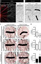
          Denervation of skeletal muscles induces severe muscle atrophy, which is preceded by cellular alterations such as increased plasma membrane permeability, reduced resting membrane potential and accelerated protein catabolism. The factors that induce these changes remain unknown. Conversely, functional recovery following denervation depends on successful reinnervation. Here, we show that activation of nicotinic acetylcholine receptors (nAChRs) by quantal release of acetylcholine (ACh) from motoneurons is sufficient to prevent changes induced by denervation. Using in vitro assays, ACh and non-hydrolysable ACh analogs repressed the expression of connexin43 and connexin45 hemichannels, which promote muscle atrophy. In co-culture studies, connexin43/45 hemichannel knockout or knockdown increased innervation of muscle fibers by dorsal root ganglion neurons. Our results show that ACh released by motoneurons exerts a hitherto unknown function independent of myofiber contraction. nAChRs and connexin hemichannels are potential molecular targets for therapeutic intervention in a variety of pathological conditions with reduced synaptic neuromuscular transmission.

          Abstract

          Denervation of muscle fibres induces muscle atrophy, via mechanisms that remain unclear. Here, the authors show that binding of acetylcoline to its receptor at the neuromuscular junction represses the expression of connexins 43 and 45, which promote atrophy, and is sufficient to prevent denervation-induced loss of myofibre mass.

          Related collections

          Most cited references57

          • Record: found
          • Abstract: found
          • Article: not found

          Intercellular calcium signaling in astrocytes via ATP release through connexin hemichannels.

          Astrocytes are capable of widespread intercellular communication via propagated increases in intracellular Ca(2+) concentration. We have used patch clamp, dye flux, ATP assay, and Ca(2+) imaging techniques to show that one mechanism for this intercellular Ca(2+) signaling in astrocytes is the release of ATP through connexin channels ("hemichannels") in individual cells. Astrocytes showed low Ca(2+)-activated whole-cell currents consistent with connexin hemichannel currents that were inhibited by the connexin channel inhibitor flufenamic acid (FFA). Astrocytes also showed molecular weight-specific influx and release of dyes, consistent with flux through connexin hemichannels. Transmembrane dye flux evoked by mechanical stimulation was potentiated by low Ca(2+) and was inhibited by FFA and Gd(3+). Mechanical stimulation also evoked release of ATP that was potentiated by low Ca(2+) and inhibited by FFA and Gd(3+). Similar whole-cell currents, transmembrane dye flux, and ATP release were observed in C6 glioma cells expressing connexin43 but were not observed in parent C6 cells. The connexin hemichannel activator quinine evoked ATP release and Ca(2+) signaling in astrocytes and in C6 cells expressing connexin43. The propagation of intercellular Ca(2+) waves in astrocytes was also potentiated by quinine and inhibited by FFA and Gd(3+). Release of ATP through connexin hemichannels represents a novel signaling pathway for intercellular communication in astrocytes and other non-excitable cells.
            Bookmark
            • Record: found
            • Abstract: found
            • Article: not found

            MIR-206 regulates connexin43 expression during skeletal muscle development

            Skeletal myoblast fusion in vitro requires the expression of connexin43 (Cx43) gap junction channels. However, gap junctions are rapidly downregulated after the initiation of myoblast fusion in vitro and in vivo. In this study we show that this downregulation is accomplished by two related microRNAs, miR-206 and miR-1, that inhibit the expression of Cx43 protein during myoblast differentiation without altering Cx43 mRNA levels. Cx43 mRNA contains two binding sites for miR-206/miR-1 in its 3′-untranslated region, both of which are required for efficient downregulation. While it has been demonstrated before that miR-1 is involved in myogenesis, in this work we show that miR-206 is also upregulated during perinatal skeletal muscle development in mice in vivo and that both miR-1 and miR-206 downregulate Cx43 expression during myoblast fusion in vitro. Proper development of singly innervated muscle fibers requires muscle contraction and NMJ terminal selection and it is hypothesized that prolonged electrical coupling via gap junctions may be detrimental to this process. This work details the mechanism by which initial downregulation of Cx43 occurs during myogenesis and highlights the tight control mechanisms that are utilized for the regulation of gap junctions during differentiation and development.
              Bookmark
              • Record: found
              • Abstract: found
              • Article: found
              Is Open Access

              Endocrine Crosstalk Between Skeletal Muscle and the Brain

              Skeletal muscle is an essential regulator of energy homeostasis and a potent coordinator of exercise-induced adaptations in other organs including the liver, fat or the brain. Skeletal muscle-initiated crosstalk with other tissues is accomplished though the secretion of myokines, protein hormones which can exert autocrine, paracrine and long-distance endocrine effects. In addition, the enhanced release or uptake of metabolites from and into contracting muscle cells, respectively, likewise can act as a powerful mediator of tissue interactions, in particular in regard to the central nervous system. The present review will discuss the current stage of knowledge regarding how exercise and the muscle secretome improve a broad range of brain functions related to vascularization, neuroplasticity, memory, sleep and mood. Even though the molecular and cellular mechanisms underlying the communication between muscle and brain is still poorly understood, physical activity represents one of the most effective strategies to reduce the prevalence and incidence of depression, cognitive, metabolic or degenerative neuronal disorders, and thus warrants further study.
                Bookmark

                Author and article information

                Contributors
                bcisterna@uc.cl
                jsaez@bio.puc.cl
                Journal
                Nat Commun
                Nat Commun
                Nature Communications
                Nature Publishing Group UK (London )
                2041-1723
                26 February 2020
                26 February 2020
                2020
                : 11
                : 1073
                Affiliations
                [1 ]ISNI 0000 0001 2157 0406, GRID grid.7870.8, Departamento de Fisiología, , Pontificia Universidad Católica de Chile, ; Santiago, Chile
                [2 ]ISNI 0000 0000 8912 4050, GRID grid.412185.b, Centro Interdisciplinario de Neurociencias de Valparaíso, Universidad de Valparaíso, ; Valparaíso, Chile
                [3 ]GRID grid.10999.38, Centro de Investigaciones Médicas, Escuela de Medicina, , Universidad de Talca, ; Talca, Chile
                [4 ]ISNI 0000 0004 6481 8274, GRID grid.499370.0, Instituto de Ciencias de la Salud, , Universidad de O’Higgins, ; Rancagua, Chile
                [5 ]GRID grid.442215.4, Facultad de Medicina y Ciencia, , Universidad San Sebastián, ; Santiago, Chile
                [6 ]GRID grid.10999.38, Thrombosis Research Center, Medical Technology School, Department of Clinical Biochemistry and Immunohaematology, Faculty of Health Sciences, , Universidad de Talca, ; Talca, Chile
                [7 ]ISNI 0000 0001 1013 7965, GRID grid.9681.6, Department of Physics, Nanoscience Center (NSC), , University of Jyväskylä, ; FI-40014 Jyväskylä, Finland
                [8 ]ISNI 0000 0001 2191 5013, GRID grid.412179.8, Centro para el Desarrollo de la Nanociencia y Nanotecnología (CEDENNA), , Universidad de Santiago de Chile, ; Santiago, Chile
                [9 ]GRID grid.441837.d, Instituto de Ciencias Biomédicas, Facultad de Ciencias de la Salud, , Universidad Autónoma de Chile, ; Santiago, Chile
                [10 ]Sección de Biología Molecular, Laboratorio Barnafi Krause, Santiago, Chile
                [11 ]GRID grid.415779.9, Sección de Biotecnología, Departamento de Salud Ambiental. Instituto de Salud Pública de Chile, ; Santiago, Chile
                [12 ]ISNI 0000 0004 0420 1184, GRID grid.274295.f, National Center for the Medical Consequences of Spinal Cord Injury, James J. Peters Veterans Affairs Medical Center, ; Bronx, NY USA
                [13 ]ISNI 0000 0001 0670 2351, GRID grid.59734.3c, Departments of Medicine and Rehabilitation Medicine, , Icahn School of Medicine at Mount Sinai, ; New York, NY USA
                Author information
                http://orcid.org/0000-0002-3421-8628
                http://orcid.org/0000-0001-6648-2135
                http://orcid.org/0000-0002-5432-9747
                http://orcid.org/0000-0001-5619-3348
                http://orcid.org/0000-0003-3811-0347
                Article
                14063
                10.1038/s41467-019-14063-8
                7044284
                32103010
                eac96895-9a6c-4432-ab87-9187f975cf54
                © The Author(s) 2020

                Open Access This article is licensed under a Creative Commons Attribution 4.0 International License, which permits use, sharing, adaptation, distribution and reproduction in any medium or format, as long as you give appropriate credit to the original author(s) and the source, provide a link to the Creative Commons license, and indicate if changes were made. The images or other third party material in this article are included in the article’s Creative Commons license, unless indicated otherwise in a credit line to the material. If material is not included in the article’s Creative Commons license and your intended use is not permitted by statutory regulation or exceeds the permitted use, you will need to obtain permission directly from the copyright holder. To view a copy of this license, visit http://creativecommons.org/licenses/by/4.0/.

                History
                : 5 September 2018
                : 10 December 2019
                Funding
                Funded by: FundRef https://doi.org/10.13039/501100002850, Fondo Nacional de Desarrollo Científico y Tecnológico (National Fund for Scientific and Technological Development);
                Award ID: 1111033
                Award ID: 1191329
                Award ID: 3170938
                Award Recipient :
                Categories
                Article
                Custom metadata
                © The Author(s) 2020

                Uncategorized
                somatic system,skeletal muscle
                Uncategorized
                somatic system, skeletal muscle

                Comments

                Comment on this article