48
views
0
recommends
+1 Recommend
0 collections
    0
    shares
      • Record: found
      • Abstract: found
      • Article: found
      Is Open Access

      Global transcriptional response of Escherichia coli O157:H7 to growth transitions in glucose minimal medium

      research-article

      Read this article at

      Bookmark
          There is no author summary for this article yet. Authors can add summaries to their articles on ScienceOpen to make them more accessible to a non-specialist audience.

          Abstract

          Background:

          Global patterns of gene expression of Escherichia coli K-12 during growth transitions have been deeply investigated, however, comparable studies of E. coli O157:H7 have not been explored, particularly with respect to factors regulating virulence genes and genomic islands specific to this pathogen. To examine the impact of growth phase on the dynamics of the transcriptome, O157:H7 Sakai strain was cultured in MOPS minimal media (0.1% glucose), RNA harvested at 10 time points from early exponential to full stationary phase, and relative gene expression was measured by co-hybridization on high-density DNA microarrays. Expression levels of 14 genes, including those encoding Shiga toxins and other virulence factors associated with the locus of enterocyte effacement (LEE), were confirmed by Q-PCR.

          Results:

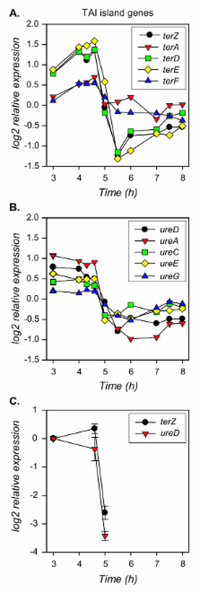
          Analysis of variance (R/MAANOVA, Fs test) identified 442 (36%) of 1239 O157-specific ORFs and 2110 (59%) of 3647 backbone ORFs that changed in expression significantly over time. QT cluster analysis placed 2468 of the 2552 significant ORFs into 12 groups; each group representing a distinct expression pattern. ORFs from the largest cluster ( n = 1078) decreased in expression from late exponential to early stationary phase: most of these ORFs are involved in functions associated with steady state growth. Also represented in this cluster are ORFs of the TAI island, encoding tellurite resistance and urease activity, which decreased ~4-fold. Most ORFs of the LEE pathogenicity island also decreased ~2-fold by early stationary phase. The ORFs encoding proteins secreted via the LEE encoded type III secretion system, such as tccP and espJ, also decreased in expression from exponential to stationary phase. Three of the clusters ( n = 154) comprised genes that are transiently upregulated at the transition into stationary phase and included genes involved in nutrient scavenging. Upregulated genes with an increase in mRNA levels from late exponential to early stationary phase belonged to one cluster ( n = 923) which includes genes involved in stress responses (e.g. gadAB, osmBC, and dps). These transcript levels remained relatively high for > 3 h in stationary phase. The Shiga toxin genes ( stx1AB and stx2B) were significantly induced after transition into stationary phase.

          Conclusion:

          Expression of more than 300 O157-specific ORFs, many implicated in virulence of the O157 pathogen, was modulated in a growth dependent manner. These results provide a baseline transcriptional profile that can be compared to patterns of gene expression of this important foodborne pathogen under adverse environmental conditions.

          Related collections

          Most cited references94

          • Record: found
          • Abstract: found
          • Article: not found

          Pathogenesis and diagnosis of Shiga toxin-producing Escherichia coli infections.

          Since their initial recognition 20 years ago, Shiga toxin-producing Escherichia coli (STEC) strains have emerged as an important cause of serious human gastrointestinal disease, which may result in life-threatening complications such as hemolytic-uremic syndrome. Food-borne outbreaks of STEC disease appear to be increasing and, when mass-produced and mass-distributed foods are concerned, can involve large numbers of people. Development of therapeutic and preventative strategies to combat STEC disease requires a thorough understanding of the mechanisms by which STEC organisms colonize the human intestinal tract and cause local and systemic pathological changes. While our knowledge remains incomplete, recent studies have improved our understanding of these processes, particularly the complex interaction between Shiga toxins and host cells, which is central to the pathogenesis of STEC disease. In addition, several putative accessory virulence factors have been identified and partly characterized. The capacity to limit the scale and severity of STEC disease is also dependent upon rapid and sensitive diagnostic procedures for analysis of human samples and suspect vehicles. The increased application of advanced molecular technologies in clinical laboratories has significantly improved our capacity to diagnose STEC infection early in the course of disease and to detect low levels of environmental contamination. This, in turn, has created a potential window of opportunity for future therapeutic intervention.
            Bookmark
            • Record: found
            • Abstract: found
            • Article: not found

            Analysis of variance for gene expression microarray data.

            Spotted cDNA microarrays are emerging as a powerful and cost-effective tool for large-scale analysis of gene expression. Microarrays can be used to measure the relative quantities of specific mRNAs in two or more tissue samples for thousands of genes simultaneously. While the power of this technology has been recognized, many open questions remain about appropriate analysis of microarray data. One question is how to make valid estimates of the relative expression for genes that are not biased by ancillary sources of variation. Recognizing that there is inherent "noise" in microarray data, how does one estimate the error variation associated with an estimated change in expression, i.e., how does one construct the error bars? We demonstrate that ANOVA methods can be used to normalize microarray data and provide estimates of changes in gene expression that are corrected for potential confounding effects. This approach establishes a framework for the general analysis and interpretation of microarray data.
              Bookmark
              • Record: found
              • Abstract: found
              • Article: not found

              Escherichia coli acid resistance: tales of an amateur acidophile.

              Gastrointestinal pathogens are faced with an extremely acidic environment. Within moments, a pathogen such as Escherichia coli O157:H7 can move from the nurturing pH 7 environment of a hamburger to the harsh pH 2 milieu of the stomach. Surprisingly, certain microorganisms that grow at neutral pH have elegantly regulated systems that enable survival during excursions into acidic environments. The best-characterized acid-resistance system is found in E. coli.
                Bookmark

                Author and article information

                Journal
                BMC Microbiol
                BMC Microbiology
                BioMed Central
                1471-2180
                2007
                29 October 2007
                : 7
                : 97
                Affiliations
                [1 ]Microbial Evolution Laboratory, National Food Safety and Toxicology Center, Michigan State University, East Lansing, Michigan 48824, USA
                Article
                1471-2180-7-97
                10.1186/1471-2180-7-97
                2241611
                17967175
                ea0c3c61-eeee-47c3-ae9f-bb1f232e88fb
                Copyright © 2007 Bergholz et al; licensee BioMed Central Ltd.

                This is an Open Access article distributed under the terms of the Creative Commons Attribution License ( http://creativecommons.org/licenses/by/2.0), which permits unrestricted use, distribution, and reproduction in any medium, provided the original work is properly cited.

                History
                : 15 April 2007
                : 29 October 2007
                Categories
                Research Article

                Microbiology & Virology
                Microbiology & Virology

                Comments

                Comment on this article