21
views
0
recommends
+1 Recommend
0 collections
    0
    shares
      • Record: found
      • Abstract: found
      • Article: found
      Is Open Access

      Heterotypic CAF-tumor spheroids promote early peritoneal metastatis of ovarian cancer

      research-article

      Read this article at

      Bookmark
          There is no author summary for this article yet. Authors can add summaries to their articles on ScienceOpen to make them more accessible to a non-specialist audience.

          Abstract

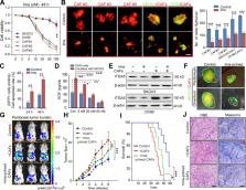
          The study provides insights in HGSOC by identifying that ascitic CAFs selectively recruit ITGA5 high ascitic tumor cells to form heterotypic spheroids named metastatic units (MUs), which actively engage in peritoneal metastasis, discriminates HGSOC from LGSOC, and act as therapeutic targets in hampering OC metastasis.

          Abstract

          High-grade serous ovarian cancer (HGSOC) is hallmarked by early onset of peritoneal dissemination, which distinguishes it from low-grade serous ovarian cancer (LGSOC). Here, we describe the aggressive nature of HGSOC ascitic tumor cells (ATCs) characterized by integrin α5 high (ITGA5 high) ATCs, which are prone to forming heterotypic spheroids with fibroblasts. We term these aggregates as metastatic units (MUs) in HGSOC for their advantageous metastatic capacity and active involvement in early peritoneal dissemination. Intriguingly, fibroblasts inside MUs support ATC survival and guide their peritoneal invasion before becoming essential components of the tumor stroma in newly formed metastases. Cancer-associated fibroblasts (CAFs) recruit ITGA5 high ATCs to form MUs, which further sustain ATC ITGA5 expression by EGF secretion. Notably, LGSOC is largely devoid of CAFs and the resultant MUs, which might explain its metastatic delay. These findings identify a specialized MU architecture that amplifies the tumor–stroma interaction and promotes transcoelomic metastasis in HGSOC, providing the basis for stromal fibroblast-oriented interventions in hampering OC peritoneal propagation.

          Graphical Abstract

          Related collections

          Most cited references45

          • Record: found
          • Abstract: found
          • Article: not found

          Stem and progenitor-like cells contribute to the aggressive behavior of human epithelial ovarian cancer.

          The cellular mechanisms underlying the increasing aggressiveness associated with ovarian cancer progression are poorly understood. Coupled with a lack of identification of specific markers that could aid early diagnoses, the disease becomes a major cause of cancer-related mortality in women. Here we present direct evidence that the aggressiveness of human ovarian cancer may be a result of transformation and dysfunction of stem cells in the ovary. A single tumorigenic clone was isolated among a mixed population of cells derived from the ascites of a patient with advanced ovarian cancer. During the course of the study, yet another clone underwent spontaneous transformation in culture, providing a model of disease progression. Both the transformed clones possess stem cell-like characteristics and differentiate to grow in an anchorage-independent manner in vitro as spheroids, although further maturation and tissue-specific differentiation was arrested. Significantly, tumors established from these clones in animal models are similar to those in the human disease in their histopathology and cell architecture. Furthermore, the tumorigenic clones, even on serial transplantation continue to establish tumors, thereby confirming their identity as tumor stem cells. These findings suggest that: (a) stem cell transformation can be the underlying cause of ovarian cancer and (b) continuing stochastic events of stem and progenitor cell transformation define the increasing aggression that is characteristically associated with the disease.
            Bookmark
            • Record: found
            • Abstract: found
            • Article: not found

            Malignant cells facilitate lung metastasis by bringing their own soil.

            Metastatic cancer cells (seeds) preferentially grow in the secondary sites with a permissive microenvironment (soil). We show that the metastatic cells can bring their own soil--stromal components including activated fibroblasts--from the primary site to the lungs. By analyzing the efferent blood from tumors, we found that viability of circulating metastatic cancer cells is higher if they are incorporated in heterotypic tumor-stroma cell fragments. Moreover, we show that these cotraveling stromal cells provide an early growth advantage to the accompanying metastatic cancer cells in the lungs. Consistent with this hypothesis, we demonstrate that partial depletion of the carcinoma-associated fibroblasts, which spontaneously spread to the lung tissue along with metastatic cancer cells, significantly decreases the number of metastases and extends survival after primary tumor resection. Finally, we show that the brain metastases from lung carcinoma and other carcinomas in patients contain carcinoma-associated fibroblasts, in contrast to primary brain tumors or normal brain tissue. Demonstration of the direct involvement of primary tumor stroma in metastasis has important conceptual and clinical implications for the colonization step in tumor progression.
              Bookmark
              • Record: found
              • Abstract: found
              • Article: not found

              A random variance model for detection of differential gene expression in small microarray experiments.

              Microarray techniques provide a valuable way of characterizing the molecular nature of disease. Unfortunately expense and limited specimen availability often lead to studies with small sample sizes. This makes accurate estimation of variability difficult, since variance estimates made on a gene by gene basis will have few degrees of freedom, and the assumption that all genes share equal variance is unlikely to be true. We propose a model by which the within gene variances are drawn from an inverse gamma distribution, whose parameters are estimated across all genes. This results in a test statistic that is a minor variation of those used in standard linear models. We demonstrate that the model assumptions are valid on experimental data, and that the model has more power than standard tests to pick up large changes in expression, while not increasing the rate of false positives. This method is incorporated into BRB-ArrayTools version 3.0 (http://linus.nci.nih.gov/BRB-ArrayTools.html). ftp://linus.nci.nih.gov/pub/techreport/RVM_supplement.pdf
                Bookmark

                Author and article information

                Journal
                J Exp Med
                J. Exp. Med
                jem
                jem
                The Journal of Experimental Medicine
                Rockefeller University Press
                0022-1007
                1540-9538
                04 March 2019
                04 March 2019
                : 216
                : 3
                : 688-703
                Affiliations
                [1 ]Cancer Biology Research Center (Key Laboratory of the Ministry of Education), Tongji Hospital, Tongji Medical College, Huazhong University of Science and Technology, Wuhan, Hubei, China
                [2 ]Department of Hematology, Tongji Hospital, Tongji Medical College, Huazhong University of Science and Technology, Wuhan, Hubei, China
                [3 ]Department of Obstetrics and Gynecology, Osaka University Graduate School of Medicine, Yamadaoka Suita, Osaka, Japan
                [4 ]Clinic of Obstetrics and Gynecology, San Gerardo Hospital, Monza, Italy
                [5 ]Department of Medicine and Surgery, University of Milan-Bicocca, Milan, Italy
                [6 ]Signaling Networks Program, Division of Oncology, Department of Medicine I, Comprehensive Cancer Center & Ludwig Boltzmann Cluster Oncology, Medical University of Vienna, Vienna, Austria
                [7 ]Department of Obstetrics and Gynecology, Experimental Tumor Immunology, University of Würzburg Medical School, Würzburg, Germany
                [8 ]Dirección de Investigación, Hospital Juárez de México D.F., Nápoles, Mexico City, Mexico
                [9 ]Division of Gynecological Oncology, NYU Langone Medical Center, Perlmutter Cancer Center, New York, NY
                [10 ]Department of Gynecological Oncology & Reproductive Medicine, University of Texas, M.D. Anderson Cancer Center, Houston, TX
                Author notes
                Correspondence to Qinglei Gao: qingleigao@ 123456hotmail.com
                [*]

                Q. Gao and Z. Yang contributed equally to this paper.

                Author information
                http://orcid.org/0000-0002-9448-3423
                http://orcid.org/0000-0001-5688-2194
                http://orcid.org/0000-0001-5461-2473
                Article
                20180765
                10.1084/jem.20180765
                6400537
                30710055
                e9c99c91-8365-457b-83c1-57917e24ed2b
                © 2019 Gao et al.

                This article is available under a Creative Commons License (Attribution 4.0 International, as described at https://creativecommons.org/licenses/by/4.0/).

                History
                : 24 April 2018
                : 02 September 2018
                : 12 October 2018
                Funding
                Funded by: National Science Foundation of China, DOI https://doi.org/10.13039/501100001809;
                Award ID: 81630060
                Award ID: 81472783
                Award ID: 81372801
                Award ID: 81572570
                Award ID: 81502250
                Award ID: 81572725
                Award ID: 81602284
                Award ID: 81772787
                Funded by: “973” Program of China
                Award ID: 2015CB553903
                Funded by: National Key Research & Development Program of China
                Award ID: 2016YFC0902901
                Funded by: National Science and Technology Major Sub-Project;
                Award ID: 2018ZX10301402-002
                Funded by: Technical Innovation Special Project of Hubei Province;
                Award ID: 2018ACA138
                Categories
                Research Articles
                Article
                307

                Medicine
                Medicine

                Comments

                Comment on this article