
- Record: found
- Abstract: not found
- Article: not found
Corneal neurotization for neurotrophic keratopathy: Review of surgical techniques and outcomes
Catherine Y. Liu ,
Andrea C. Arteaga ,
Sammie E. Fung ,
M. Soledad Cortina ,
Ilya M. Leyngold ,
Vinay K. Aakalu
April 2021
April 2021
There is no author summary for this article yet. Authors can add summaries to their articles on ScienceOpen to make them more accessible to a non-specialist audience.
Related collections
Most cited references84
- Record: found
- Abstract: found
- Article: not found
Evaluation and management of peripheral nerve injury.
William W Campbell (2008)
- Record: found
- Abstract: found
- Article: not found
Nerve physiology: mechanisms of injury and recovery.
Ron Menorca, Theron Fussell, John C Elfar (2013)
- Record: found
- Abstract: found
- Article: found
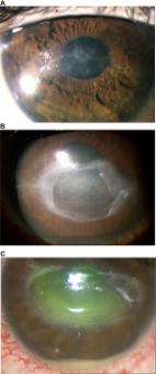
Diagnosis and management of neurotrophic keratitis
Marta Sacchetti, Alessandro Lambiase (2014)
Author and article information
Comments
Comment on this article
scite_
0
0
0
0
0
0
0
0
Citing PublicationsSupportingMentioningContrasting
See how this article has been cited at scite.ai
scite shows how a scientific paper has been cited by providing the context of the citation, a classification describing whether it supports, mentions, or contrasts the cited claim, and a label indicating in which section the citation was made.