9
views
0
recommends
+1 Recommend
0 collections
    0
    shares
      • Record: found
      • Abstract: found
      • Article: not found

      Metabolic reprogramming by the S-nitroso-CoA Reductase system protects from kidney injury

      research-article

      Read this article at

      ScienceOpenPublisherPMC
      Bookmark
          There is no author summary for this article yet. Authors can add summaries to their articles on ScienceOpen to make them more accessible to a non-specialist audience.

          Abstract

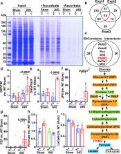
          Endothelial nitric oxide (NO) synthase (eNOS) is protective against kidney injury, but the molecular mechanisms are poorly understood 1, 2 . NO-based cellular signaling is generally mediated by protein S-nitrosylation, the oxidative modification of Cys residues to form S-nitrosothiols (SNOs). S-nitrosylation regulates proteins in all functional classes, and is controlled by enzymatic machinery including S-nitrosylases and denitrosylases that add and remove SNO from proteins, respectively 3, 4 . We recently reported in Saccharomyces cerevisiae that the classic metabolic intermediate Co-enzymeA (CoA) serves as an endogenous source of SNOs through its conjugation with NO to form S-nitroso-CoA (SNO-CoA), and that S-nitrosylation of proteins by SNO-CoA is governed by its cognate denitrosylase, SNO-CoA reductase (SCoR) 5 . Mammals possess a functional homologue of yeast SCoR, an aldo-keto reductase family member (AKR1A1) 5 with an unknown physiological role. Here we report that the SNO-CoA/AKR1A1 (SCoR) system is highly expressed in renal proximal tubules where it transduces the activity of eNOS in reprogramming intermediary metabolism, thereby protecting kidneys from acute kidney injury (AKI). Specifically, AKR1A1 deletion in mice to reduce SCoR activity increased protein S-nitrosylation, protected against AKI and improved survival, whereas renoprotection was lost in Akr1a1 −/− / eNOS −/− mice. Metabolic profiling coupled with unbiased mass spectrometry-based SNO-protein identification revealed that protection by the SNO-CoA/SCoR system is mediated by inhibitory S-nitrosylation of pyruvate kinase M2 (PKM2) through a novel locus of regulation, thereby balancing fuel utilization (through glycolysis) with redox protection (through the pentose phosphate shunt). Targeted deletion of PKM2 from mouse proximal tubules recapitulated precisely the protective and mechanistic effects of S-nitrosylation in Akr1a1 −/− mice, whereas Cys-mutant PKM2 refractory to S-nitrosylation negated SNO-CoA bioactivity. Our discoveries provide a first physiological function of the SNO-CoA/SCoR system in mammals, reveal novel regulation of renal metabolism and of PKM2 in differentiated tissues in particular, and offer a new perspective on kidney injury with therapeutic implications.

          Related collections

          Most cited references37

          • Record: found
          • Abstract: found
          • Article: not found

          Pathophysiology of ischemic acute kidney injury.

          Acute kidney injury (AKI) as a consequence of ischemia is a common clinical event leading to unacceptably high morbidity and mortality, development of chronic kidney disease (CKD), and transition from pre-existing CKD to end-stage renal disease. Data indicate a close interaction between the many cell types involved in the pathophysiology of ischemic AKI, which has critical implications for the treatment of this condition. Inflammation seems to be the common factor that links the various cell types involved in this process. In this Review, we describe the interactions between these cells and their response to injury following ischemia. We relate these events to patients who are at high risk of AKI, and highlight the characteristics that might predispose these patients to injury. We also discuss how therapy targeting specific cell types can minimize the initial and subsequent injury following ischemia, thereby limiting the extent of acute changes and, hopefully, long-term structural and functional alterations to the kidney.
            Bookmark
            • Record: found
            • Abstract: found
            • Article: not found

            Pyruvate kinase M2 activators promote tetramer formation and suppress tumorigenesis

            Cancer cells engage in a metabolic program to enhance biosynthesis and support cell proliferation. The regulatory properties of pyruvate kinase M2 (PKM2) influence altered glucose metabolism in cancer. PKM2 interaction with phosphotyrosine-containing proteins inhibits enzyme activity and increases availability of glycolytic metabolites to support cell proliferation. This suggests that high pyruvate kinase activity may suppress tumor growth. We show that expression of PKM1, the pyruvate kinase isoform with high constitutive activity, or exposure to published small molecule PKM2 activators inhibit growth of xenograft tumors. Structural studies reveal that small molecule activators bind PKM2 at the subunit interaction interface, a site distinct from that of the endogenous activator fructose-1,6-bisphosphate (FBP). However, unlike FBP, binding of activators to PKM2 promotes a constitutively active enzyme state that is resistant to inhibition by tyrosine-phosphorylated proteins. These data support the notion that small molecule activation of PKM2 can interfere with anabolic metabolism.
              Bookmark
              • Record: found
              • Abstract: found
              • Article: not found

              Regulation of pyruvate metabolism and human disease

              Pyruvate is a keystone molecule critical for numerous aspects of eukaryotic and human metabolism. Pyruvate is the end-product of glycolysis, is derived from additional sources in the cellular cytoplasm, and is ultimately destined for transport into mitochondria as a master fuel input undergirding citric acid cycle carbon flux. In mitochondria, pyruvate drives ATP production by oxidative phosphorylation and multiple biosynthetic pathways intersecting the citric acid cycle. Mitochondrial pyruvate metabolism is regulated by many enzymes, including the recently discovered mitochondria pyruvate carrier, pyruvate dehydrogenase, and pyruvate carboxylase, to modulate overall pyruvate carbon flux. Mutations in any of the genes encoding for proteins regulating pyruvate metabolism may lead to disease. Numerous cases have been described. Aberrant pyruvate metabolism plays an especially prominent role in cancer, heart failure, and neurodegeneration. Because most major diseases involve aberrant metabolism, understanding and exploiting pyruvate carbon flux may yield novel treatments that enhance human health.
                Bookmark

                Author and article information

                Journal
                0410462
                6011
                Nature
                Nature
                Nature
                0028-0836
                1476-4687
                16 November 2018
                28 November 2018
                January 2019
                28 May 2019
                : 565
                : 7737
                : 96-100
                Affiliations
                [1 ]Institute for Transformative Molecular Medicine and Department of Medicine, Case Western Reserve University and University Hospitals Cleveland Medical Center, Cleveland, Ohio 44106, USA,
                [2 ]Center for Proteomics and Bioinformatics, Case Western Reserve University School of Medicine, Cleveland, Ohio 44106, USA.
                [3 ]Nephrology and Endocrine Divisions, Massachusetts General Hospital and Harvard Medical School, Boston, Massachusetts 02114, USA.
                [4 ]Broad Institute of MIT and Harvard, Cambridge, Massachusetts 02139, USA.
                [5 ]Division of Nephrology and Department of Medicine, Beth Israel Deaconess Medical Center and Harvard Medical School, Boston, Massachusetts 02215, USA.
                [6 ]Center for Vascular Biology Research, Beth Israel Deaconess Medical Center and Harvard Medical School, Boston, Massachusetts 02215, USA.
                [7 ]Department of Medicine, Cedars-Sinai Medical Center, Los Angeles, California, 90048, USA
                [8 ]Harrington Discovery Institute, University Hospitals Cleveland Medical Center, Cleveland, Ohio 44106, USA.
                Author notes

                Author contributions

                H.Z. and J.S.S. designed the study. H.Z. carried out most of experiments and analyzed the results. R.Z. performed AKI surgery. P.A. prepared samples for iTRAQ LC-MS/MS and metabolomics. C.T.S. prepared samples for photolysis-chemiluminescence assay. A.H. and P.A. purified SCoR from bovine kidney. Z.Q. handled mice. L.W. performed quantitative iTRAQ LC-MS/MS. E. R. and S. M. P. contributed to project conception and carried out metabolomics analyses. S.A.K. contributed to project conception and performed histological stains. H.Z. and J.S.S. wrote the manuscript with input from all authors.

                [* ]Materials & Correspondence: Institute for Transformative Molecular Medicine, Case Western Reserve University, Wolstein Research Building 4129, 2103 Cornell Road, Cleveland, OH 44106, Tel.: 216-368-5725, Fax: 216-368-2968, jonathan.stamler@ 123456case.edu
                Correspondence and requests for materials should be addressed to J.S.S ( jonathan.stamler@ 123456case.edu ).
                Article
                NIHMS1511348
                10.1038/s41586-018-0749-z
                6318002
                30487609
                e7077c56-4fad-4ec1-ba40-766c77f011e0

                Users may view, print, copy, and download text and data-mine the content in such documents, for the purposes of academic research, subject always to the full Conditions of use: http://www.nature.com/authors/editorial_policies/license.html#terms

                Reprint and permissions information is available at www.nature.com/reprints. Readers are welcome to comment on the online version of the paper.

                History
                Categories
                Article

                Uncategorized
                Uncategorized

                Comments

                Comment on this article